
Image
|
Figure Caption
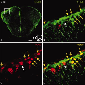
Fig. 4
Some proliferating cells at the ventricle of the lesioned hemisphere express the radial glial marker S100B. A: Overview of a section through the site of injury. The injured hemisphere (to the left) expresses higher levels of S100B and PCNA (see Fig. 3A). B–D: High magnification view of boxed region in A. PCNA positive cells co-express S100B (yellow arrows in B–D). Some clusters of PCNA positive cells express only weak levels of S100B (white arrow in B–D) or no S100B (red arrow in B–D). Scale bar = 100 μm in A; 10 μm in B–D.
Acknowledgments
This image is the copyrighted work of the attributed author or publisher, and
ZFIN has permission only to display this image to its users.
Additional permissions should be obtained from the applicable author or publisher of the image.
Full text @ Dev. Dyn.