
|
Fig. 3
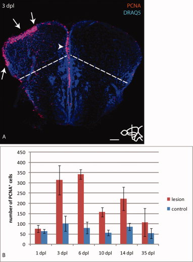
Stab injury leads to up-regulation of PCNA in the lesioned hemisphere. A: Immunostaining for the proliferation marker PCNA combined with a nuclear stain (Draq5) at 3 dpl. Arrows indicate up-regulation of PCNA at the ventricle of the lesioned hemisphere, mainly restricted to the pallium delineated by a dashed line. The arrowhead indicates proliferating cells at the dorso-medial ventricle of the lesioned hemisphere closely adjacent to the control hemisphere. B: Quantification of PCNA positive cells at the pallial ventricle of the lesioned hemisphere compared with the control hemisphere shows normal proliferation levels at 1 dpl, a peak of proliferation at the lesioned side between 3 and 6 dpl and significantly increased levels until at least 14 dpl. At 35 dpl PCNA levels are comparable between lesioned and control hemispheres. n=5 telencephala per time-point. Error bars indicate standard deviation. Scale bar = 100 μm in A.