
|
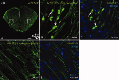
Fig. 2
GFAP-GFP positive radial glial fibers exhibit hypertrophic swellings in response to stab injury. A: Overview of a GFAP-GFP brain section through the lesioned area. Boxes indicate regions shown in higher magnification in B, C (left box) and D, E (right box). B,C: High magnification of the boxed region in A, with arrows indicating that cell body-like swellings do not co-stain with the nuclear marker Draq5 (C), suggesting that they are not parenchymal GFAP-GFP positive cells (B is an average projection of the confocal Z-stack to illustrate fiber morphology). D,E: No hypertrophic swelling can be found in the unlesioned hemisphere, suggesting a reaction specific to injury (D is an average projection of the confocal Z-stack to illustrate fiber morphology). Scale bar = 100 μm in A; 7.5 μm in B–E.