
|
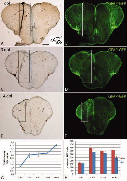
Fig. 1
Stab injury of the telencephalon leads to up-regulation of GFAP-GFP and swelling of radial glial processes. All views of this and following figures are transverse sections through the telencephalon, stained with the markers indicated (color-coded). Inserts indicate rostro-caudal positions. Lesioned telencephalic hemispheres are always located to the left. A–F: Brightfield images of cross-sections through lesioned areas. Boxes indicate lesion (A,C,E). Immunostaining against GFP in the Tg(GFAP-GFP) transgenic line at different time-points after injury (B,D,F). A slight up-regulation can be detected at 3 dpl (C,D), which is increased at 14 dpl (E,F). Boxes indicate region of injury. G: Measurement of GFAP-GFP fluorescent signal intensity using ImageJ and calculating the fold-change between injured and uninjured hemisphere shows increasing GFAP-GFP intensity from 3 dpl until 14 dpl. H: Quantification of S100B positive cells comparing lesioned to unlesioned hemispheres at different time points post lesion. There is no significant change in S100B positive cells until 14 dpl, when an increase in S100B cells can be observed. Scale bar = 100 μm in A–F.