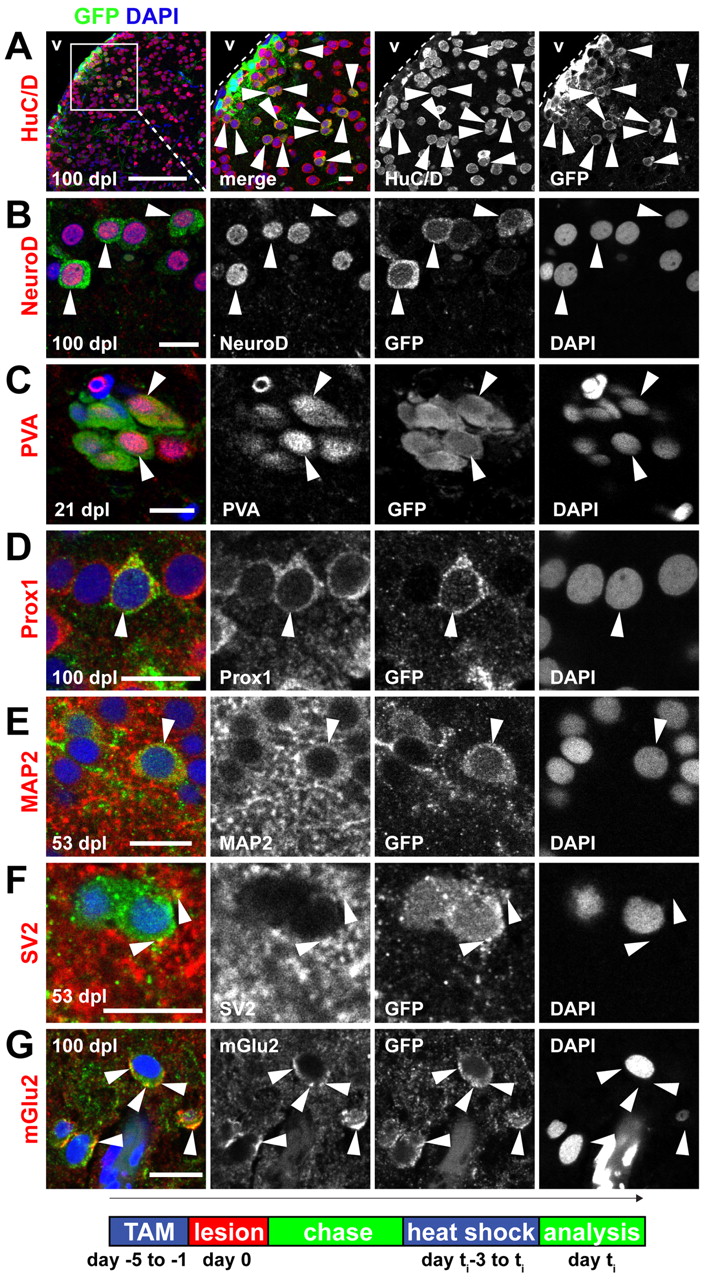
Fig. 7 Radial glia-derived neurons are maintained long term, show a correct regional identity and display features of mature functional neurons. (A) Many recombined GFP+(green)/HuC/D+(red) neurons (arrowheads) are maintained in the parenchyma and in the PVZ until 100 dpl. (B-G) Examples of radial glia-derived (GFP+, green) neurons in the dorsal parenchyma that express: (B) the dorsal marker NeuroD1 (arrowheads, red) at 100 dpl; (C) the mature interneuron and regional marker parvalbumin (arrowheads, PVA, red) at 21 dpl; (D) the regional marker Prox1 (arrowhead, red) at 100 dpl; (E) the mature neuronal marker MAP2a+b (arrowhead, MAP2, red) at 53 dpl; (F) the synaptic vesicle marker SV2 (arrowheads, SV2, red) at 53 dpl; and (G) the synaptic marker metabotropic glutamate receptor 2 (arrowheads, mGlu2, red) at 100 dpl. Scale bars: 100 μm in A overview; 10 μm in A inset; in 10 μm in B-G. v, ventricle. Timeline shows experimental procedures.
Image
Figure Caption
Acknowledgments
This image is the copyrighted work of the attributed author or publisher, and
ZFIN has permission only to display this image to its users.
Additional permissions should be obtained from the applicable author or publisher of the image.
Full text @ Development

