
Fig. 4
- ID
- ZDB-IMAGE-111031-5
- Publication
- Kroehne et al., 2011 - Regeneration of the adult zebrafish brain from neurogenic radial glia-type progenitors
- All Figures
- Figures for Kroehne et al., 2011
|
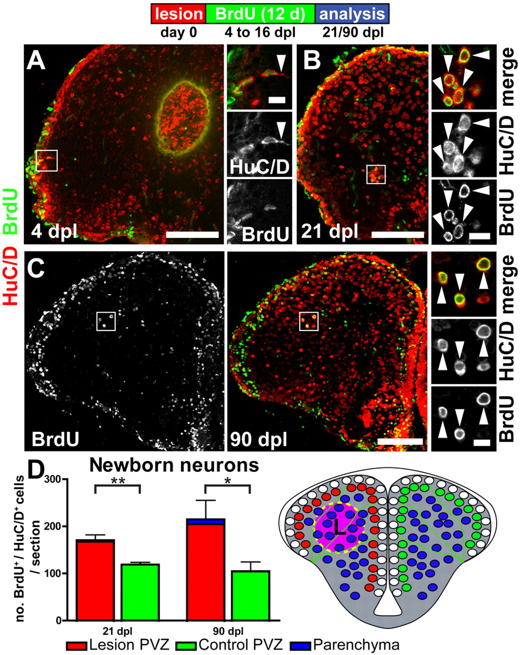
Fig. 4 Reactive neurogenesis leads to many long-lived, newly generated neurons. (A) 4 dpl newborn BrdU+(green)/HuC/D+(red) neuroblasts, generated at the time of the radial glia proliferation peak (BrdU pulse 48-92 hpl), are leaving the VZ and migrate towards the lesion site. Note the prominent leading edge profile (arrowhead), a typical feature of migrating neuroblasts. (B) Newborn BrdU+/HuC/D+ neurons that were born between 4 and 16 dpl can be found close to the lesion site in the parenchyma 21 dpl (arrowheads). (C) Many of these injury-induced ectopic neurons survive for at least 90 dpl close to the lesion site deep in the parenchyma, as well as in the PVZ, as shown by BrdU/HuC/D double staining (arrowheads). Scale bars: 100 μm in A-C; 10 μm in insets. (D) Quantification of BrdU+/HuC/D+ cells shows a significant net gain of newly generated neurons in the lesioned hemisphere at 21 (n=5) and 90 dpl (n=3). Data are mean+s.e.m. **Pd0.01; *Pd0.05.