
Image
|
Figure Caption
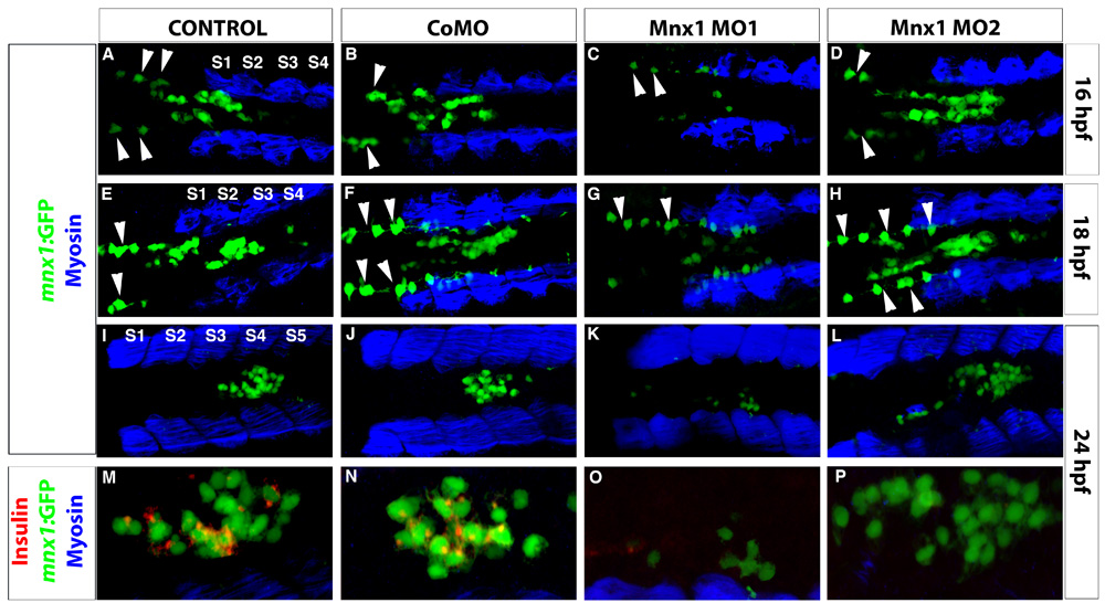
Fig. S9 Mnx1 MO1 but not Mnx1 MO2 disrupts Tg(mnx1:GFP) expression. (A-P) Confocal images of Tg(mnx1:GFP) embryos at (A-D) 16 hpf, (E-H) 18 hpf and (I-P) 24 hpf. (A-L) Immunolabeling with GFP and myosin antibodies. Endodermal Tg(mnx1:GFP) cells (green) are observed in the midline adjacent to somites (S) (blue). Dorsally localized GFP-expressing cells anterior to the somites are motoneurons (arrowheads). (M-P) Higher magnifications. (I-L) Colabeling of GFP cells with insulin antibody (red).
Acknowledgments
This image is the copyrighted work of the attributed author or publisher, and
ZFIN has permission only to display this image to its users.
Additional permissions should be obtained from the applicable author or publisher of the image.
Full text @ Development