
|
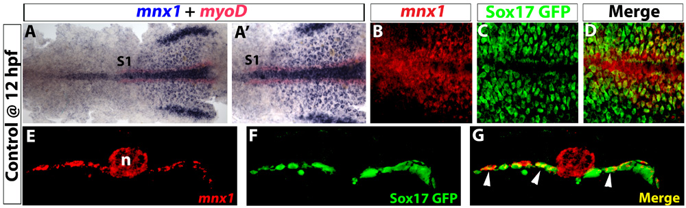
Fig. S1 Early expression of mnx1 is localized in pre-pancreatic endoderm. (A-D) Ventral view of 12 hpf Tg(sox17:EGFP) embryos. (A) In situ hybridization for mnx1 (blue) and myoD (red). (A′) High-magnification view. (B-D) Confocal images of mnx1 transcripts (fluorescent images of chromogenic in situ hybridizations, see Material and methods) in A,A′ (B) and GFP immunostaining (C); colocalization of mnx1 transcripts and GFP is observed (D). (E-G) Vibratome section (100 µm) taken through the level of somite 1 (see A,A′). Confocal z-stacks of mnx1 expression (E), GFP labeling (F) and merged images (G) are shown to demonstrate endodermal expression of mnx1 (arrowheads in G). Anterior to the left. S1, somite 1; n, notochord.