
Fig. 2
- ID
- ZDB-IMAGE-111025-4
- Genes
- Antibodies
- Publication
- Perlin et al., 2011 - Neuronal Neuregulin 1 type III directs Schwann cell migration
- All Figures
- Figures for Perlin et al., 2011
|
Fig. 2
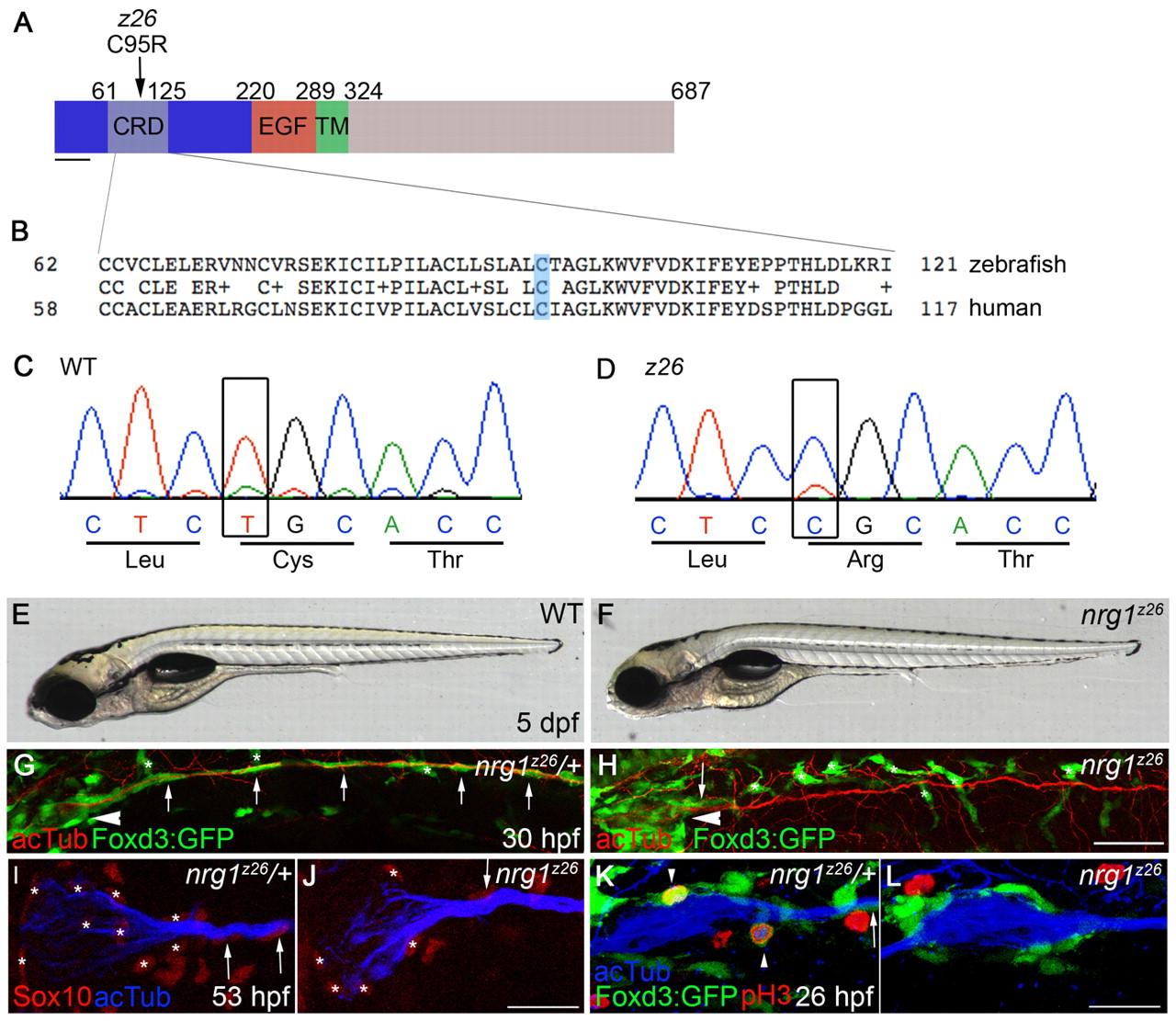
nrg1z26 specifically disrupts the Nrg1 type III isoform. (A) Protein structure of zebrafish Nrg1 type III. The z26 lesion in the cysteine-rich domain (CRD) of the type III-specific domain (dark blue) changes a conserved cysteine (C) at position 95 to an arginine (R). TM, transmembrane domain. Scale bar: 40 amino acids. (B) Comparison of zebrafish and human Nrg1 type III CRD sequences. The conserved cysteine mutated by the z26 lesion is shaded in blue. (C,D) Sequence traces from wild-type and z26 mutant zebrafish larvae illustrating the T-to-C lesion. (E,F) Lateral views of 5 dpf wild-type and nrg1z26 mutant sibling embryos. (G,H) Lateral views of 30 hpf nrg1z26/+ siblings (G) and nrg1z26 mutants (H) stained with anti-acetylated tubulin (acTub, red). Arrowheads mark the PLLg, asterisks mark pigment cells. Embryos were genotyped after imaging. (I,J) PLLg of a 53 hpf nrg1z26/+ sibling (I) and nrg1z26 mutant (J) stained with anti-Sox10 (red) and anti-acTub (blue). Asterisks mark Schwann cells associated with the ganglion. (K,L) PLLg of 26 hpf nrg1z26/+ sibling (K) and nrg1z26 mutant (L) stained with anti-phospho-Histone H3 (pH3) (red) and anti-acTub (blue). Arrowhead marks Foxd3:GFP+ cells that are co-labeled with pH3. (G-L) Schwann cells are marked by Foxd3:GFP (green, arrows). Anterior is to left, dorsal is up. (I-L) Embryos were genotyped before imaging and images are a projection of either three (I,J) or four (K,L) 3 μm sections taken at the middle of each PLLg. Scale bars: 50 μm in G,H; 25 μm in I-L.