
IMAGE
Fig. S4
- ID
- ZDB-IMAGE-111025-35
- Publication
- Clark et al., 2011 - Generation of Rab-based transgenic lines for in vivo studies of endosome biology in zebrafish
- All Figures
- Figures for Clark et al., 2011
Image
|
Figure Caption
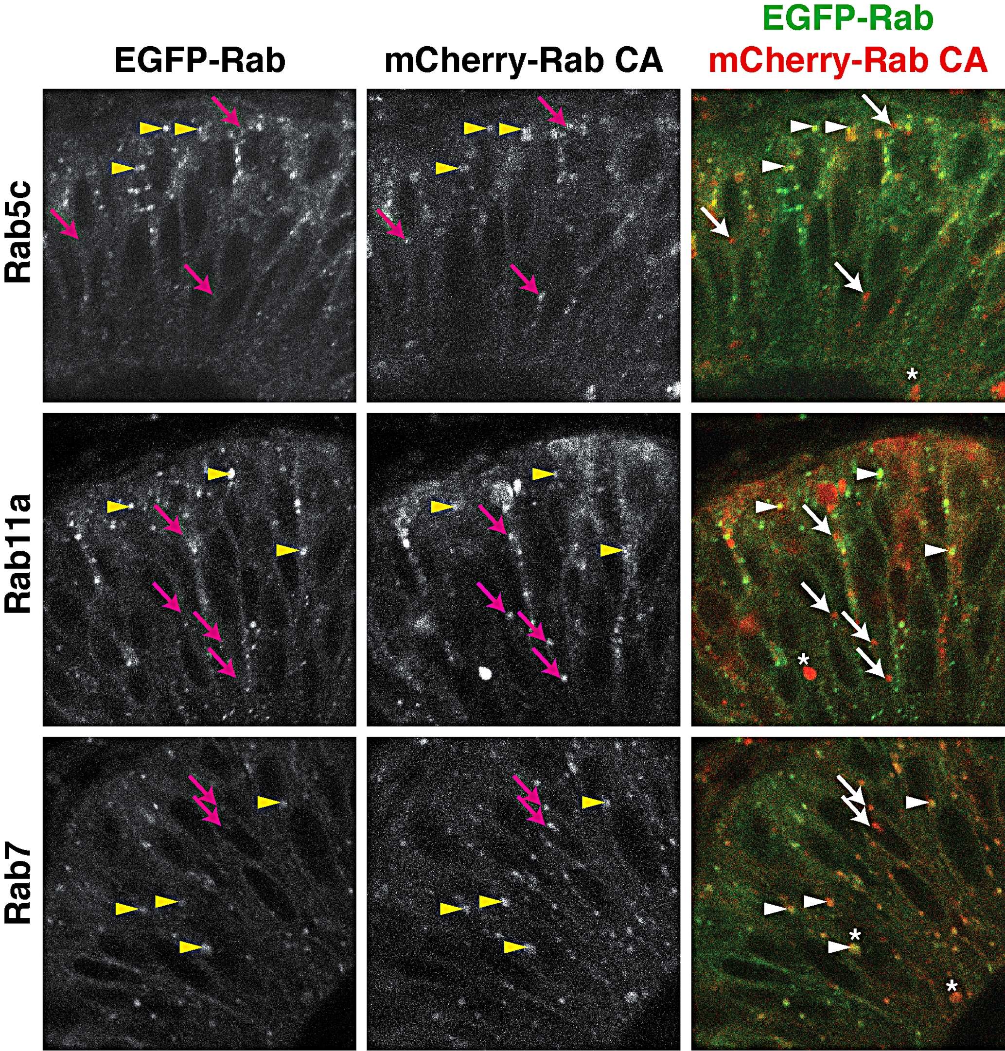
Fig. S4
Partial co-localization of EGFP-Rab and mCherry-Rab consitutive active proteins. Fifty-micrometer, single-plane, confocal sections of retinal neuroepithelial cells from EGFP-Rab transgenics injected with corresponding mCherry-rab CA mRNA (for example, EGFP-Rab5c embryos injected with mCherry-rab5c CA mRNA). Arrowheads indicate mCherry-RabCA puncta that co-localize with transgenic EGFP-Rab vesicles, whereas arrows indicate non-colocalizing mCherry-RabCA puncta. Asterisks illustrate the presence of large vesicular structures that undergo dynamic movements in time-lapse images similar to EGFP-Rab vesicles.
Acknowledgments
This image is the copyrighted work of the attributed author or publisher, and
ZFIN has permission only to display this image to its users.
Additional permissions should be obtained from the applicable author or publisher of the image.
Full text @ Dev. Dyn.