
Fig. S4
- ID
- ZDB-IMAGE-111025-12
- Publication
- Perlin et al., 2011 - Neuronal Neuregulin 1 type III directs Schwann cell migration
- All Figures
- Figures for Perlin et al., 2011
|
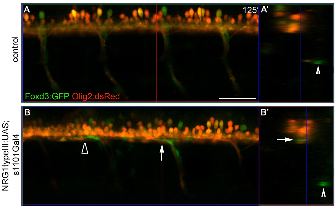
Fig. S4
Schwann cells migrate within the spinal cord of embryos overexpressing human NRG1 type III. (A,B) Lateral views of single scans from in vivo time-lapse movies of a control embryo (A; see Movie 1 in the supplementary material) and an embryo overexpressing human NRG1 type III in all neurons (B; see Movie 2 in the supplementary material) at 125 minutes. Schwann cells are marked by the Foxd3:GFP transgene (green) and oligodendrocytes and motoneurons are marked by Olig2:dsRed (red). (A2,B2) Orthogonal view of the yz plane at the location of the vertical red lines in A and B. Arrowhead marks PLLn (A2,B2); arrow points to Foxd3:GFP staining within the Olig2:dsRed-marked spinal cord (B2). The open arrowhead marks the Schwann cell that is tracked by the open arrowhead in Fig. 6D-I,K,K2. Anterior is to the left, dorsal is up. Comparable regions over the yolk extension were imaged. Embryos were genotyped after imaging. Scale bar: 50 μm.