
|
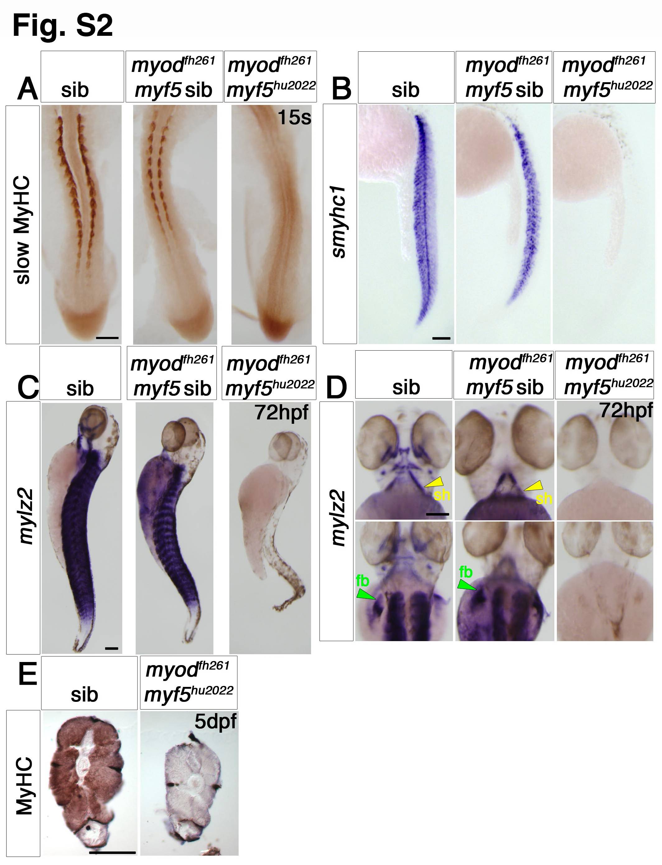
Fig. S2 Myf5 or Myod is required for myogenesis. Dorsal flatmounts (A,D—bottom panel), ventral (D—top panel) and lateral (B,C) wholemounts or transverse cryosections (E) of zebrafish embryos from myf5hu2022/+;myodfh261/+ in-cross analysed at the indicated stage by immunohistochemistry for slow MyHC (A) or all MyHC (E) or in situ mRNA hybridization for smyhc1 (B) or mylz2 (C,D), anterior to top (A–D), dorsal to top (E). A. At 15 s, most embryos show strong slow muscle differentiation, but approximately 3/16ths (genotyped as myodfh261/fh261;myf5hu2022/+ or myodfh261/fh261;myf5+/+) have less muscle and 1/16th (genotyped as myodfh261/fh261;myf5hu2022/hu2022) have no detectable muscle. B. mRNAs encoding slow myosin is reduced in putative myodfh261 mutants and absent in doubles. C,D. At 72 hpf, no mylz2 mRNA is detected in genotyped double mutants, in which the trunk and tail are severely reduced in size. E. At 120 hpf, double mutant lacks MyHC in the gut extension region and has a reduced somite area. Note the similar sizes of non-muscle tissues. Bars = 100 μm.
Reprinted from Developmental Biology, 358(1), Hinits, Y., Williams, V.C., Sweetman, D., Donn, T.M., Ma, T.P., Moens, C.B., and Hughes, S.M., Defective cranial skeletal development, larval lethality and haploinsufficiency in Myod mutant zebrafish, 102-12, Copyright (2011) with permission from Elsevier. Full text @ Dev. Biol.