
|
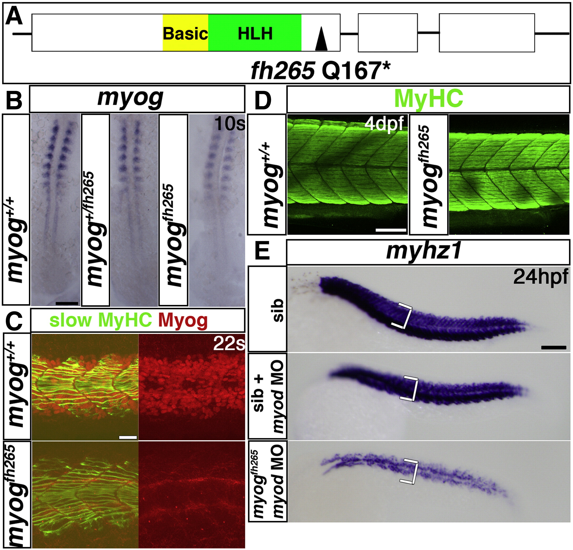
Fig. 2 Myogenin cooperates with Myod in fast myogenesis. A. Schematic of Myog gene and protein showing the fh265 glutamine to stop mutation following the helix–loop–helix domain. B–E. In situ mRNA hybridization for myog (B) or myhz1 (E) or immunodetection of Myog and slow MyHC (C) or MyHC (D) in myog+/fh265 incrosses, injected with myod morpholino as indicated (E). B. Expression of myog mRNA is little affected in myogfh265 mutants. Dorsal flatmounts, anterior to top. C. Nuclear immunoreactivity with a polyclonal anti-rat Myog antibody is lost in myogfh265 mutants at 22 s. D. All embryos from a myog+/fh265 incross show normal levels of MyHC at 4 dpf. E. No change in myhz1 mRNA is detected in myogfh265 mutants at 24 hpf (top panel). Injection of myod MO into myogfh265 (bottom panel) leads to a greater loss of fast muscle than when injected into their siblings (middle panel), which are comparable to myodfh261 mutants (see Fig. 1F). Lateral views, anterior to left (C–E). Bars = 100 μm (except C = 20 μm).
Reprinted from Developmental Biology, 358(1), Hinits, Y., Williams, V.C., Sweetman, D., Donn, T.M., Ma, T.P., Moens, C.B., and Hughes, S.M., Defective cranial skeletal development, larval lethality and haploinsufficiency in Myod mutant zebrafish, 102-12, Copyright (2011) with permission from Elsevier. Full text @ Dev. Biol.