
Fig. 4
- ID
- ZDB-IMAGE-111018-20
- Publication
- Mouillesseaux et al., 2011 - Mutation in utp15 Disrupts Vascular Patterning in a p53-Dependent Manner in Zebrafish Embryos
- All Figures
- Figures for Mouillesseaux et al., 2011
|
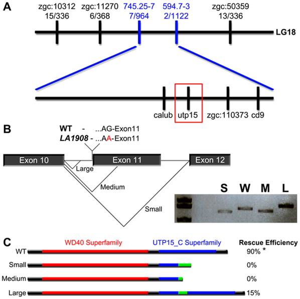
Fig. 4 The LA1908 locus encodes zebrafish utp15.
A, Positional cloning showed tight linkage between LA1908 and utp15. B, Sequence analysis of the utp15 gene revealed an intronic G to A point mutation, abolishing the splice acceptor site preceding exon11 and resulting in three utp15 cryptic splicing variants; a 134 bp deletion (utp15 small), a 26 bp deletion (utp15 medium), and a 36 bp insertion (utp15 large). Inset in (B) shows the relative cDNA sizes of S (utp15 small), W (wild type utp15), M (utp15 medium), and L (utp15 large) variants. C, The protein structure and translational consequences of utp15 splice variants. Utp15 small and medium proteins contain premature stop codons resulting in truncated proteins. Injecting in vitro synthesized mRNA generated from these constructs was unable to rescue LA1908 mutant phenotype. Utp15 large contains a 12 amino acid insertion and displays some ability to rescue phenotype. Red bar, WD40 protein-protein interaction domain. Blue bar, utp15 C-terminal superfamily domain of unknown function. Green bar, altered peptide sequence resulting from cryptic splicing. * = p-value <0.001 by Chi-Squared analysis of Mendelian ratios of a minimum of 100 embryos per condition. Rescue efficiency is calculated as # embryos exhibiting LA1908 mutant phenotype/# total embryos with LA1908 mutant genotype, n = 10 (WT), 28 (small), 11 (medium), and 13 (large) mRNA injected mutant embryos.