
|
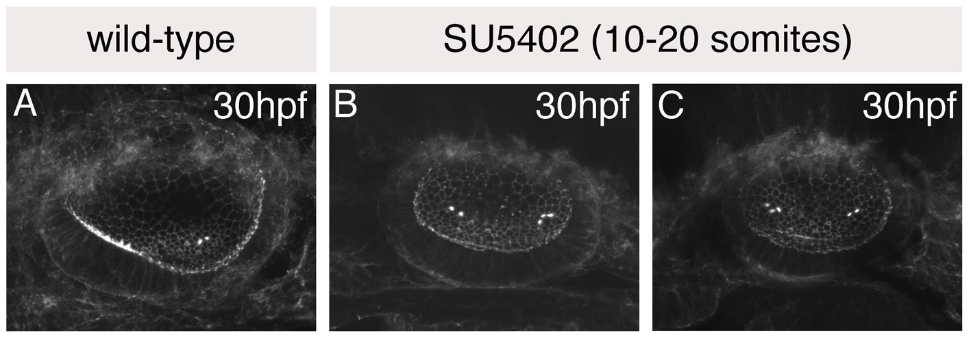
Fig. S1 The single medial macula in SU5402-treated otic vesicles originates as two separate groups of hair cells. (A-C) Otic vesicles stained with FITC-phalloidin to reveal the actin-rich stereociliary bundles of sensory hair cells. (A) Wild-type (DMSO-treated) control, showing anterior hair cells in a ventral position, and posterior hair cells in a medial position. (B,C) SU5402-treated embryos treated with 10 μM SU5402 for 5 hours from 10 to 20S; two examples are shown. Two groups of hair cells (towards the anterior and posterior of the vesicle) are detected in SU5402-treated otic vesicles, as in controls; however, these are both positioned medially, resembling the posterior patch in control embryos. Lateral views; anterior to the left.