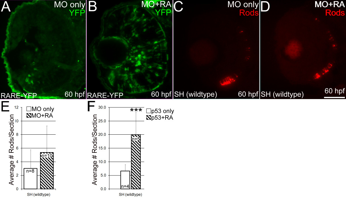
Fig. 12 The effects of exogenous retinoic acid on RARαb morphants. (A, B) RARE-YPF embryos injected with rarαb/p53 MO and fixed at 60 hpf. YFP expression was detected on 4 μm cryosections using an anti-GFP antibody. (A) RARE-YFP embryos injected with the rarαb/p53 MO show reduced expression of YFP in the ventral retina. (B) RARE-YFP embryo injected with rarαb/p53 MO and treated with RA beginning at 36 hpf. Upregulation of YFP expression is evident in cells across all the retinal layers. (C, D) Wildtype (SH) embryos injected with rarαb/p53 MO and fixed at 60 hpf, sectioned at 4 μm, and labeled with an antibody to rod opsin (1D1). (C) Embryo injected only with rarαb/p53 MO. (D) Embryo injected with rarαb/p53 MO and treated with RA beginning at 36 hpf. (E) Numbers of rod photoreceptors were quantified by counting them on sections (see Methods). Statistically significant differences between DMSO and RA-treated groups were not detected in wildtype RARαb morphants. (DMSO group n = 8, average 9 sections per eye; RA group n = 9, average 8 sections per eye) (n = 13, average 8 sections per eye). Bar = 50 μm. (F) Numbers of rod photoreceptors per section in embryos treated with the p53 MO only, and exposed to DMSO (n = 4, average 9 sections per eye) or RA (n = 5, average 8 sections per eye), and examined at 63 hpf. In this experiment RA treatment results in significantly higher numbers of rods. Bar = 50 μm.
Image
Figure Caption
Acknowledgments
This image is the copyrighted work of the attributed author or publisher, and
ZFIN has permission only to display this image to its users.
Additional permissions should be obtained from the applicable author or publisher of the image.
Full text @ BMC Dev. Biol.

