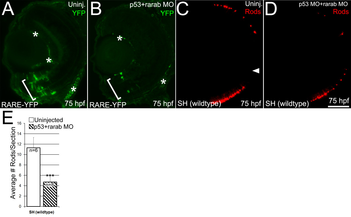
Fig. 11 Endogenous retinoic acid signaling and production of rods is reduced by knockdown of RARαb expression. (A, B) RARE-YFP embryos at 75 hpf. YFP expression was detected on embryos sectioned at 4 μm using an anti-GFP antibody. (A) In uninjected RARE-YFP embryos, there is robust expression of YFP in cells in the ventral retina (bracket). Non-specific antibody staining is indicated by asterisks. (B) RARE-YFP embryos injected with the rarαb/p53 MO show a marked reduction in the number of cells expressing YFP in the ventral retina (bracket). (C, D) Wildtype (SH) embryos at 75 hpf. (C) Tissue section from uninjected wildtype embryo labeled with a rod opsin antibody (1D1). Arrowhead: optic nerve head. (D) Injection of the rarαb/p53 MO into wildtype embryos results in reduced expression of 1D1 rod opsin antigen. (E) Numbers of rod photoreceptors were quantified by counting them on sections (see Methods). Wildtype embryos injected with the rarαb/p53 MO (n = 8, average 8 sections per eye) at 75 hpf had a significant reduction in the number of rods per section compared to uninjected embryos (n = 6, average 10 sections per eye); p < 0.5, Student′s T-Test). Bar = 50 μm.
Image
Figure Caption
Acknowledgments
This image is the copyrighted work of the attributed author or publisher, and
ZFIN has permission only to display this image to its users.
Additional permissions should be obtained from the applicable author or publisher of the image.
Full text @ BMC Dev. Biol.

