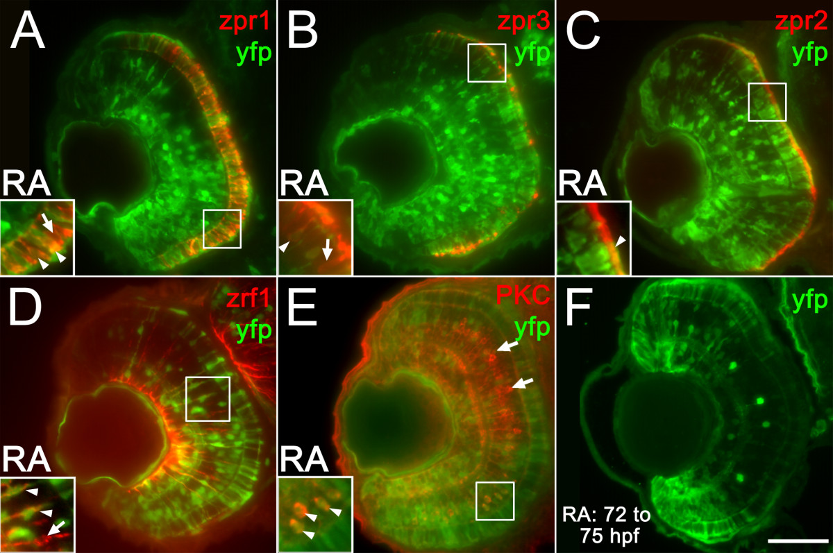
Fig. 9 Multiple retinal cell types engage in retinoic acid signaling in response to retinoic acid treatment during retinal differentiation. (A to E) Embryos carrying the RARE-YFP transgene were treated with 0.3 μM RA at 48 hpf, and at 75 hpf were processed as 5 μm cryosections for indirect immunofluorescence with an anti-GFP antibody (green color in all panels) and the following markers (red color in all panels): zpr1 (stains red and green-sensitive cones; (A), zpr3 (stains rods and green cones; (B), zpr2 (stains RPE; C), zrf1 (stains Müller glia; D), and anti-PKC (stains rod bipolar cells; E). Dorsal is up in all panels; boxed regions in each panel appear at higher magnification in the insets. All types of retinal cells examined are capable of responding directly to RA by activating the RARE-YFP transgene (doubly-labeled cells in each panel; arrowheads; colabeling within the limit of resolution of our objective lens = 1.4 μm), although not all cells of each type respond (singly-labeled cells; arrows). (F) Embryos carrying the RARE-YFP transgene were treated with 0.3 μM RA at 72 hpf, and were fixed at 75 hpf as 5 μm cryosections for indirect immunofluorescence with the anti-GFP antibody. Widespread transgene expression indicates rapid response to RA. Bar = 50 μm.
Image
Figure Caption
Acknowledgments
This image is the copyrighted work of the attributed author or publisher, and
ZFIN has permission only to display this image to its users.
Additional permissions should be obtained from the applicable author or publisher of the image.
Full text @ BMC Dev. Biol.

