
|
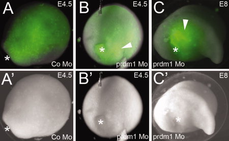
Fig. 4
prdm1 Mo never incorporates in ectoderm. A–C: Fluorescent images showing incorporation of fluorescein isothiocyanate (FITC) -labeled morpholino (green). A2–C2: Brightfield images of the same embryos. A,A2: Embryonic day (E) 4.5 embryo injected with a control morpholino, showing incorporation of the morpholino throughout the dorsal ectoderm. The embryo is shown in dorsal view, with the anterior in the top right-hand corner. Asterisk denotes the position of the blastopore. B,B2: E4.5 embryo injected with prdm1 morpholino. Morpholino is incorporated in the mesoderm and endoderm (white arrowhead). The embryo is shown with the dorsal side facing up, and the posterior (blastopore, marked with an asterisk) facing toward the viewer. C,C2:prdm1 morpholino-injected embryo at E8. Orientation: the head is facing right, and the dorsal side is facing up. Asterisk denotes the abnormal position of the blastopore.