Fig. 5
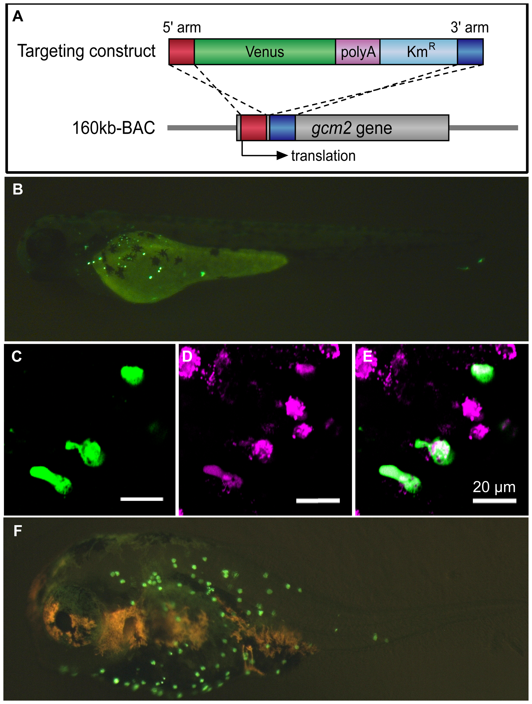
Analysis of gcm2 enhancers specific for ionocytes on the skin surface of zebrafish.
(A) Construction of the 160 kb BAC-Venus. The targeting construct was amplified by PCR from a plasmid containing Venus, polyA, and Kmr. Each primer contained 50 bp of the gcm2-derived sequences that served as homology arms for homologous recombination. The 160 kb BAC contained sequences that were 120 kb upstream and 40 kb downstream of the gcm2 locus. After homologous recombination, Venus was inserted into the translation site (160 kb BAC-Venus). (B) A 160 kb BAC-Venus transient transgenic zebrafish (72 hpf). Venus-expressing cells are observed on the skin surface but not in the gills. (C) Several cells on the yolk sac expressed Venus. (D) Ionocytes stained with MitoTracker. (E) Merge of (C) and (D), showing overlap in staining with a subset of ionocytes. (F) A 160 kb BAC-Venus transient transgenic 7-day-old fugu. Venus-expressing cells are observed on the skin surface.

