
|
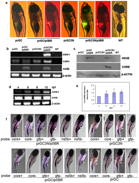
Fig. 2
Amplification of the HCV sub-replicon in liver of the zebrafish larvae.
a. HCV sub-replicon amplified in those co-injected with prGC3N and p5BR vectors. Green fluorescence represents the NS5B-dependent HCV core protein products of the sub-replicon (prGC3N/p5BR); red fluorescence indicates the presence of the NS5B enzyme. Arrows point the positive signals in liver area. WT, untreated control. b. RT-PCR measurement for the amplification of the study genes in the sub-replicon. Core+, the positive strand of HCV core RNA; core-, the negative strand of HCV core RNA; ns5b, HCV ns5b from p5BR; and beta-actin, loading control. WT, wild type. c. Western blot for HCV core and NS5B proteins with anti-core or anti-NS5B antibody, respectively. Beta-actin was loaded as a control. d. Amplification of the HCV sub-replicon exhibits a time–dependent increase from 4- to 10-dpf in zebrafish larvae for the positive strand of core RNA; negative strand core RNA and beta-actin remains to be constant. dpf, day post fertilization. e. Confirmation of the time–dependent amplification of the HCV sub-replicon with quantitative real time RT-PCR for the positive strand of HCV core RNA. f. Whole mount in situ hybridization of the co-injected zebrafish. Whole mount in situ hybridizations were carried out on 10-dpf larvae using antisense or sense RNA probes. The presented are original microscopy images of the zebrafish larvae (80X). dpf, day post fertilization.