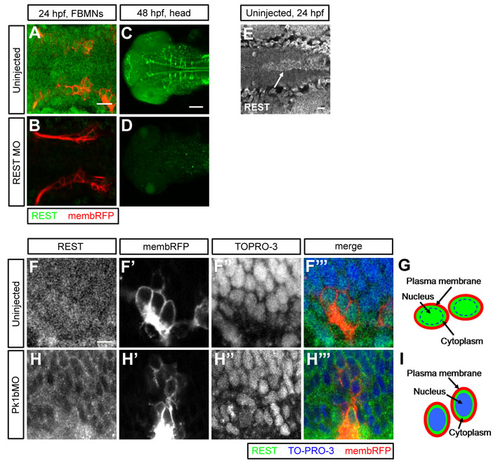
Fig. S6 Immunohistochemistry reveals localization of endogenous REST protein. (A-D) Dorsal views of 24 hpf (A,B) and 48 hpf (C,D) Tg(zCREST1:membRFP) embryos. Uninjected (A,C) and REST morphant (B,D) embryos were immunostained for REST (green). FBMNs are visible in A and B (red). REST expression is elevated in several subsets of cells, including reticulospinal neurons (C). Full-length REST protein levels are substantially reduced in REST morphants. E) Dorsal view of the hindbrain in a 24 hpf embryo immunostained for REST. Arrow highlights elevated REST expression in the floor plate. (F-F′′,H-H′′) Single-slice dorsal views of FBMNs (red) in 24 hpf uninjected (F-F′′) and Pk1b morphant (H-H′′) Tg(zCREST1:membRFP) embryos immunostained for REST (green), with nuclei labeled by TO-PRO-3 (blue). (G,I) REST localizes throughout the cell body of wild-type FBMNs, including in the nuclei (G). In Pk1b morphants, REST is reduced in the nuclei and enriched in the cytoplasm (I).
Image
Figure Caption
Figure Data
Acknowledgments
This image is the copyrighted work of the attributed author or publisher, and
ZFIN has permission only to display this image to its users.
Additional permissions should be obtained from the applicable author or publisher of the image.
Full text @ Development

