
Fig. 3
- ID
- ZDB-IMAGE-110720-47
- Publication
- Patil et al., 2011 - Functional Analysis of Retinitis Pigmentosa 2 (RP2) Protein Reveals Variable Pathogenic Potential of Disease-Associated Missense Variants
- All Figures
- Figures for Patil et al., 2011
|
Fig. 3
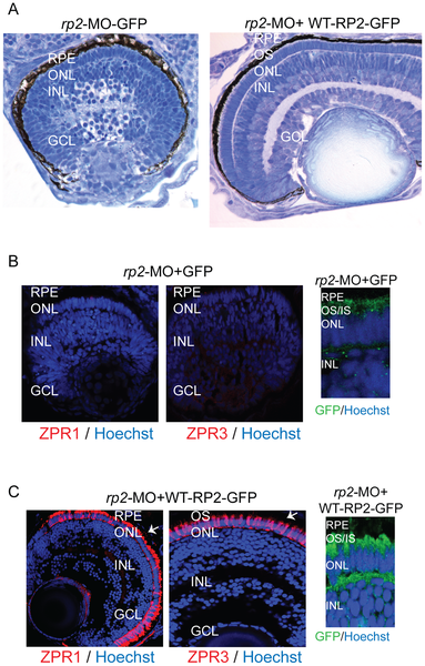
Human WT RP2 can rescue rp2-MO associated phenotype.
A. Histological analysis of retinas from embryos co-injected with rp2-MO and mRNA encoding GFP (left panel; rp2-MO + GFP) or wild type (WT) RP2-GFP fusion protein (right panel; rp2-MO + RP2-GFP). OS: outer segment; IS: inner segment; ONL: outer nuclear layer; OPL: outer plexiform layer. B and C. Immunofluorescence analysis of the indicated embryo retinas was performed using Zpr1 or Zpr3 antibodies (red; arrows) or anti-GFP antibodies (green). Hoechst was used to stain nuclei (blue). OS: outer segment; IS: inner segment; ONL: outer nuclear layer; INL: inner nuclear layer; GCL: ganglion cell layer.