
|
Fig. 4
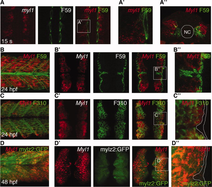
Fibre-type specific expression of myl1. A: Expression of myl1 (red) was detected in the fast domain of the paraxial mesoderm in a 15-somite-stage flat-mounted embryo using fluorescent in situ hybridisation. Labelling of adaxial cells/slow fibres using F59 antibody (green) showed no overlap with the myl1 staining. A2: Blow-up of a single somite as indicated by square in A. A3: Transverse section of 15-somite-stage embryo. NC, notochord. Labelling of adaxial cells/slow fibres using F59 antibody (green) showed no overlap with the myl1 staining. B: Lateral view of 24-hpf embryo stained with F59 antibody (green), labelling slow fibers and myl1 antisense probe (red). B2: Transverse sections of 24-hpf embryo where slow fibres, detected with F59 antibody (green), showed no co-localisation with myl1 antisense probe (red). B3: Blow-up of region boxed in B2. C: Lateral view of 24-hpf embryo where F310 antibody (green) labelling fast fibres is detected in myl1-positive fibres (red). C2: Transverse sections of 24-hpf embryo where fast fibres detected with F310 antibody (green) co-localised with myl1 antisense probe (red). C3: Blow-up of region boxed in C2. D: Lateral view of 48-hpf Tg(mylz2:GFP)i135 transgenic embryo where GFP+ fast fibres were co-labelled with myl1 antisense probe (red). D2: Transverse sections of 48-hpf Tg(mylz2:GFP)i135 transgenic embryo where GFP+ fast fibres were co-labelled with myl1 antisense probe (red). D3: Blow-up of region boxed in D2.