
|
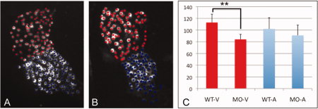
Fig. 7
The tbx2ab morphant has fewer ventricular cardiomyocytes by 2 dpf. A,B: Shown are representative hearts from wild-type (A) or tbx2ab morphant (B) embryos derived from the myl7:dsRed-nuc reporter line. Flat-mounted embryos were chosen that displayed a clear constriction at the position of the presumptive AVC and were imaged by confocal microscopy. C: Based on this morphological distinction, individual cells were marked as ventricular (red) or atrial (blue) and quantified as shown in the chart. For each sample n = 4, and this was reproducible evaluating independent batches of embryos. The morphant ventricle (but not the atrium) has significantly fewer cardiomyocytes (indicated on the Y-axis) compared with wild-type (P < 0.01, according to Students t-test).