
Fig. 5
- ID
- ZDB-IMAGE-110712-53
- Publication
- Sedletcaia et al., 2011 - Heart chamber size in zebrafish is regulated redundantly by duplicated tbx2 genes
- All Figures
- Figures for Sedletcaia et al., 2011
|
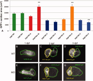
Fig. 5
Chamber sizes are altered in the tbx2ab morphants. Hearts were imaged in wild-type or tbx2ab double morphant embryos and the area of atrial and ventricular chambers was outlined and measured. A: The top panel shows the quantification from representative embryos at 1 days postfertilization (dpf), 2 dpf, or 3 dpf as indicated. Also as indicated, the measurement was in wild-type (WT) or morphant (MO), either in the heart tube (1 dpf, green), or for the atrium (A) or ventricle (V). In each case n is at least 10. B–F: Lower panels show representative images of wild-type and tbx2ab morphant hearts at 1 dpf (B,E), 2 dpf (C,F) and 3 dpf (D,G). In B and E the heart tube is outlined in green. In C, D, F, and G, yellow outlines the ventricle and green outlines the atrium. The ** indicates statistical significance compared with wild-type, according to Student′s t-test, P < 0.01.