
|
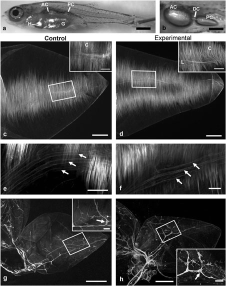
Fig. 6
Anatomy of larval swimbladders from control (n = 8) and experimental (n = 8) groups at the LDC stage of development. a: Representative larva from control group (total body length 9.5 mm) showing anterior (AC) and posterior (PC) chambers of swimbladder in relation to the gut (G; partly obscuring swimbladder) and heart (H). b: Higher magnification image of swimbladder in situ in opened coelomic cavity; AC and PC connected by a narrowed ductus communicans (DC). Panels c, e, g show images of tissues from control group; d, f, h show images of tissue from experimental group. c, d: Phalloidin labeled smooth muscle fibers of lateral muscle bands in the wall of the PC in both groups. Boxes in main panels show locations of insets, which depict circular (C) and longitudinal (L) muscle fibers. e, f: Smooth muscle was associated with blood vessels of the rete (arrows), as demonstrated by phalloidin labeling in the wall of craniolateral portion of the PC in tissue from both groups. g, h: Zn-12 immunoreactivity showed robust neuronal staining within the walls of both swimbladder chambers and in nerves coursing to the swimbladder along the pneumatic duct (arrowhead). In each panel, insets show details of regions indicated by boxes; putative neuronal cell bodies were observed in swimbladders from both control and experimental groups (arrows). Scale bars represent: a, 1 mm; b, 0.5 mm; c, d, 100 μm (insets, 50 μm); e, f, 50 μm; g, h, 200 ^μm (insets, 50 μm).