
|
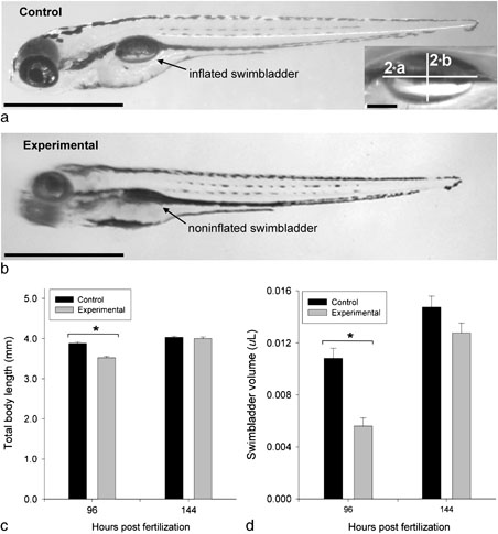
Fig. 2
Effects of SMG on swimbladder morphology, total body length, and swimbladder volume during development. a, b: Representative photomicrographs of left lateral views of larvae with inflated swimbladders from control (a) and larvae with uninflated swimbladders from experimental (b) groups at 96 hpf. Inset shows magnified view of single-chambered swimbladder with the long (2*a) and short (2*b) axes marked for volumetric analysis, as modeled by a rotary prolate ellipsoid (see text). In b, the arrow marks the position of the uninflated swimbladder in larvae exposed to SMG. In all images of swimbladder in this and subsequent figures, cranial is to the left and dorsal is upward. c: Plots of total body length of larvae from control (black bars: n = 42) and experimental (gray bars: n = 70) groups measured at 96 and 144 hpf (control: n = 17; experimental: n = 18). d: Plots of swimbladder volumes estimated from animals that had inflated swimbladders at 96 hpf (control: n = 26; experimental: n = 10) and 144 hpf (control: n = 17; experimental: n = 18) with bar shading as in c. Asterisk (*) indicates a significant difference (independent samples t-test, P<0.001) in mean values between groups at the same time point. In panels a and b, scale bars represent 1.0 mm; in inset, 100 μm.