
Image
|
Figure Caption
Fig. 7
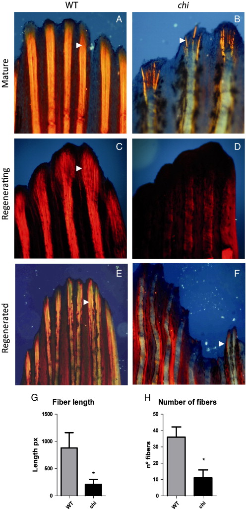
col1a1a dominant chi/+ mutant shows aberrant fin exoskeleton. Actinotrichia and lepidotrichia pattern in mature (A–B, E–F) and regenerating fin at 4 dpa (C–D). Wild type (A, C and E) and heterozygous chihuahua mutant (col1a1adcl24/+; B, D and F). Arrowhead represents actinotrichia. (G–H) Statistical comparisons of length and number of actinotrichia per fin ray between chi mutant (black column) and wild type (gray column). All images were obtained under polarized optics following staining with Picrosirius red. Asterisks represent significant difference by Mann–Whitney u-test.
Figure Data
Acknowledgments
This image is the copyrighted work of the attributed author or publisher, and
ZFIN has permission only to display this image to its users.
Additional permissions should be obtained from the applicable author or publisher of the image.
Reprinted from Developmental Biology, 354(1), Durán, I., Marí-Beffa, M., Santamaría, J.A., Becerra, J., and Santos-Ruiz, L., Actinotrichia collagens and their role in fin formation, 160-172, Copyright (2011) with permission from Elsevier. Full text @ Dev. Biol.