
|
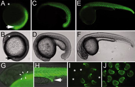
Fig. 2
EGFP fluorescence profile of Tg(wnt8aPAC:EGFP) heterozygotes. A,B: Expression at bud stage, lateral view, anterior to upper left. Arrow indicates tailbud, asterisk indicates anterior neural plate. Note strong fluorescence limited to the posterior embryo. C,D: Expression at <18-somite stage, lateral view, anterior to left. Fluorescence is observed in somites with an increasing gradient toward the tailbud. E,F: Expression at 24 hpf, lateral view. Strong fluorescence continues in somitic mesoderm, with continuing fluorescence detectable around the yolk. Expression is also observed in the heart (G, outlined by arrows) and pronephros (H, arrow). I,J: Comparison of transgene expression in embryos derived from males or females. I: Progeny from a heterozygous male crossed to wild-type female. Fifty percent of embryos fluoresce, 50% do not (non-fluorescing embryos indicated with asterisks). Note easily scored expression in somites of trunk and tail and around yolk. J: In contrast, 100% of progeny from a heterozygous female crossed to a wild-type male fluoresce throughout all tissues.