Fig. s2
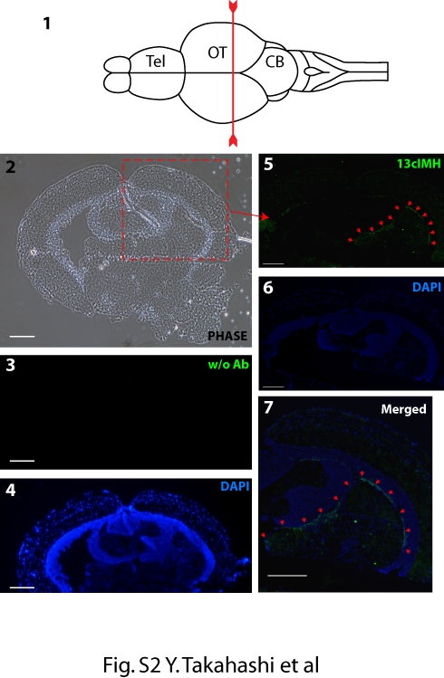
Immunohistochemistry of the cross section at the optic tectum of zebrafish brain. (1) The diagram shows zebrafish brain (modified from Castro, A. et al. [3]) and a line indicates the cutting line. Tel; Telencephalon, OT; Optic Tectum CB; Cerebellum. (2) A phase contrast image of a cross section of zebrafish brain. (3, 4) The brain section was incubated without the monoclonal antibody for 13cIMH (Negative control, 3; FITC channel, 4; DAPI). (5-7) The brain section was incubated with the monoclonal antibody for 13cIMH. Green fluorescence signals, indicated by red arrows, for 13cIMH (5; 13cIMH, 6; DAPI, 7; merged) were detected in the preventricular grey zone (PGZ) of optic tectum (OT). Scale bar = 200 μm.

