
|
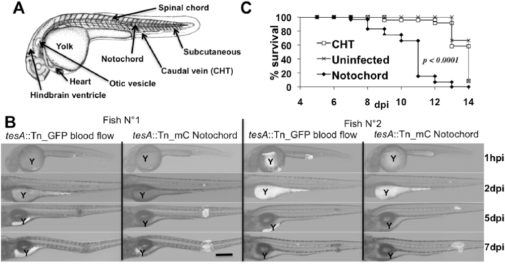
Fig. 6
Notochordal infections. A. Camera lucida sketch of the 31 h embryo (http://zfin.org). Injection sites are indicated. B. Individual embryos where sequentially injected at 30 hpf with the tesA::Tn_GFP strain in the blood flow and the tesA::Tn_mCherry strain in the notochord. Embryos were individually imaged to follow infection. Two representative embryos are depicted as overlays of phase contrast plus GFP fluorescence or RFP fluorescence at, respectively, 1 hpi, 2, 5 and 7 dpi. Both embryos died at 8 dpi. Scale bar is 250 μm. Autofluorescence of the yolk (Y) is seen in both GFP and mCherry channels. C. Survival curves of notochord-infected embryos. Approximately 93 ± 49 (three replicates, mean ± SD) cfu tesA::Tn mutants were injected at 30 hpf in the CHT (n = 24) or in the notochord (n = 59). Death was monitored on a daily basis.