
|
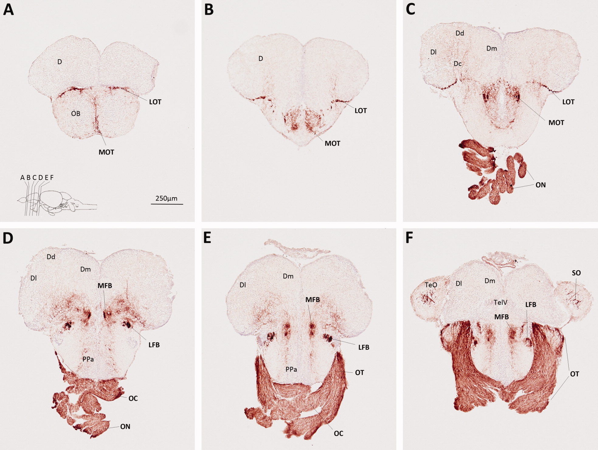
Fig. 3
Myelin P0 expression in the telencephalon, optic nerves, and adjacent structures. Transverse sections of adult zebrafish brain, from posterior olfactory bulbs to anterior tectum, were labeled using P0 primary antibody, and a histochemical reaction yielding a red product. A blue nuclear counterstain was used to facilitate identification of anatomical features. The sections, which are oriented dorsal upwards, progress in a rostrocaudal direction; their planes are indicated in the inset to A, which also shows the scale bar for all six panels. Anatomical landmarks are indicated on the left side of each image, and P0-immunoreactive structures are labeled in bold on the right side of each image. For anatomical annotations, please see list of abbreviations.