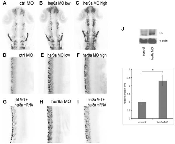
Fig. 5 Her8a morphants exhibit upregulation of HuC/D expression.
HuC/D expression is upregulated in Her8a morpholino injected embryos analyzed by immunohistochemistry with anti-Hu antibody at 24 hpf. The black-and-white fluorescent signals were inverted to negative film for a better presentation. (A and D) embryos injected with control morpholino. (B and E) 6 ng of MO1 or 4 ng of MO2 injection (low dose) resulted in indistinguishable phonotype and therefore only embryos injected with 4 ng of MO2 are shown (see Fig. S2 for MO1). (C and F) Injection of 12 ng MO1 or 8 ng MO2 (high dose) resulted in identical morphological defects and only MO2 injected embryos are shown, which shows more dramatic upregulation of HuC/D expression. (A–C) Brain regions; (D-I) 3-somite to 9-somite level of the spinal cord. (A–F) Hu-positve cells were significantly increased in the morpholino injected embryos. (G–I) The phenotype can be rescued by co-injection of morpholino with her8a mRNA. (J) Western blot analysis confirming the levels of HuC/D expression in Her8a morphants were up-regulated in comparison to the control. * p<0.05.

