
Fig. 8
- ID
- ZDB-IMAGE-110607-6
- Publication
- Hami et al., 2011 - Zebrafish cardiac development requires a conserved secondary heart field
- All Figures
- Figures for Hami et al., 2011
|
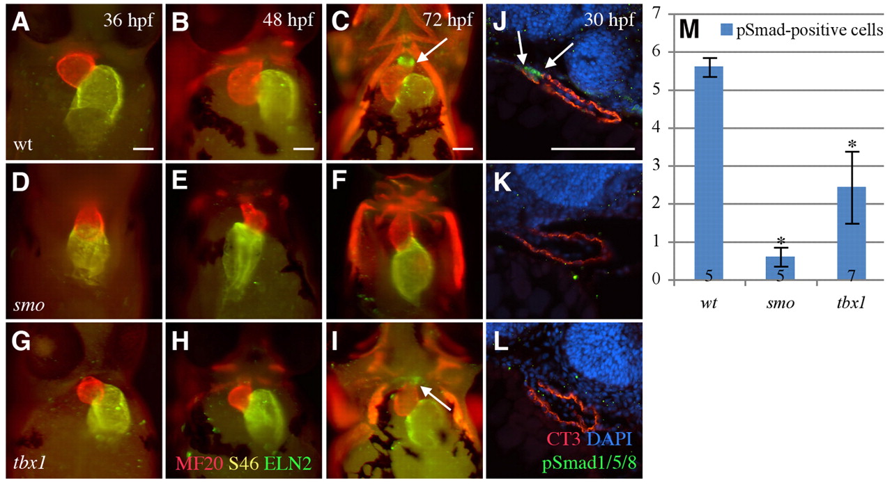
Fig. 8 Zebrafish tbx1 and smo mutants display arterial pole defects. (A-C) Ventral view of whole-mount wild-type zebrafish, with cranial to the top. The heart is looped by 36 hpf and the bulbus arteriosus (Eln2) is formed by 72 hpf. Ventricle (MF20, red); atrium (S46, yellow). (D-I) In smo (D-F) and tbx1 (G-I) mutants, cardiac development is impaired. (J) Sagittal sections from the left show pSmad1/5/8 (green and arrows) near the arterial pole of the wild-type heart (CT3) at 30 hpf. (K,L) In the smo (K) and tbx1 (L) mutants, pSmad1/5/8 is significantly reduced. (M) Quantification of pSmad1/5/8-positive cells in wild-type versus smo and tbx1 mutants. Numbers in bars indicate the number of fish sampled. *, P=1.23×10⁻³. Error bars indicate s.d. Scale bars: 5 μm.