
Fig. 4
- ID
- ZDB-IMAGE-110607-3
- Publication
- Hami et al., 2011 - Zebrafish cardiac development requires a conserved secondary heart field
- All Figures
- Figures for Hami et al., 2011
|
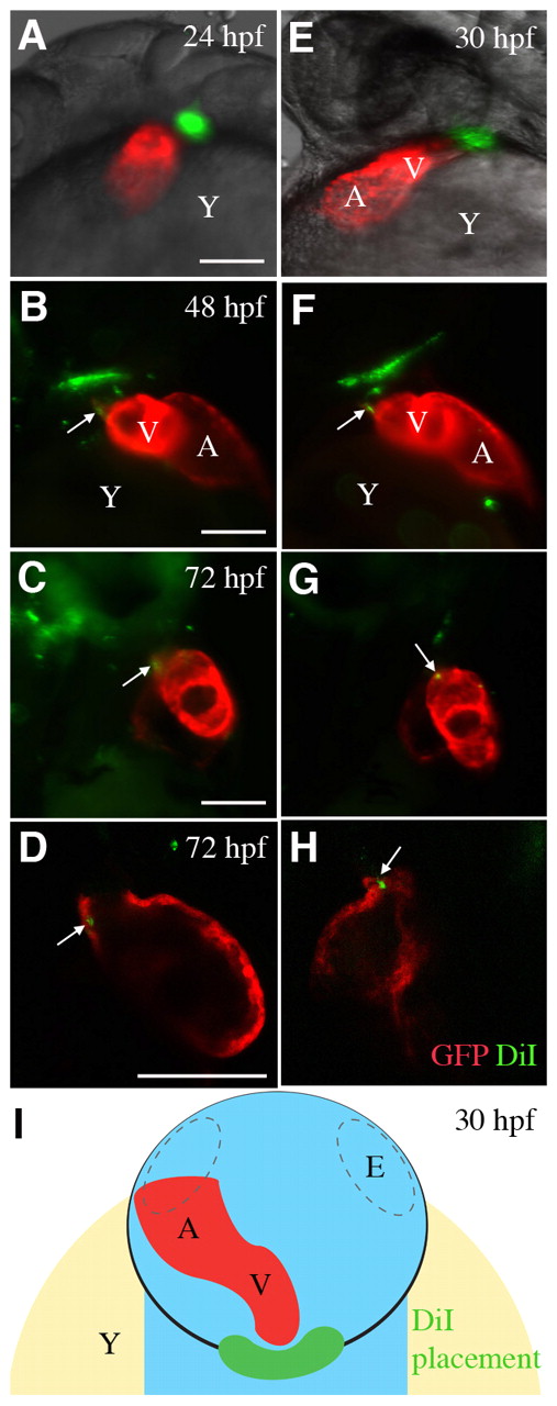
Fig. 4 Cells in the pericardial wall are incorporated into the developing heart tube. (A,E) Cells were labeled with DiI (pseudocolored green) adjacent to the arterial pole of the heart tube (pseudocolored red) in Tg(cmlc2::GFP) zebrafish at (A) 24 hpf and (E) 30 hpf. Pseudocoloring improved the visibility of DiI-labeled cells. Zebrafish are shown from the left side, cranial to the top. (B,C,F,G) Labeled cells were observed in the GFP-positive myocardium at (B,F) 48 hpf and (C,G) 72 hpf. Zebrafish are shown from the right side, cranial to the top. (D,H) Optical sections of 72 hpf zebrafish hearts from the right side, cranial to the top, confirming that DiI-labeled cells were incorporated into the myocardium. (I) Diagram showing the approximate area of labeled cells (green) in the pericardial wall (black line) when viewed dorsally at 30 hpf. The heart tube is shown (red). Dashed lines indicate eyes (E). A, atrium; V, ventricle; Y, yolk. Scale bars: 10 μm.