
IMAGE
Fig. 1
- ID
- ZDB-IMAGE-110527-28
- Publication
- Hong et al., 2011 - The transcriptional mediator component med12 is required for hindbrain boundary formation
- All Figures
- Figures for Hong et al., 2011
Image
|
Figure Caption
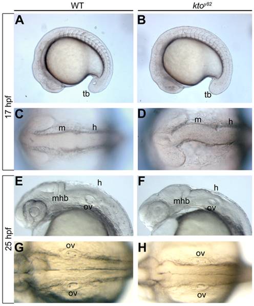
Fig. 1 The hindbrain phenotype of ktoy82.
Live images of lateral (A,B,E,F) and dorsal views (C, D, G, H) of the developing hindbrain at 17 hpf (A–D) and 25 hpf (E–H). The early midbrain and hindbrain regions are malformed at 17 hpf in mutant embryos (D), and no ventricle is visible at 25 hpf (H). h, hindbrain; m, midbrain; mhb, mid-hindbrain boundary; ov, otic vesicle; tb, tail bud.
Figure Data
Acknowledgments
This image is the copyrighted work of the attributed author or publisher, and
ZFIN has permission only to display this image to its users.
Additional permissions should be obtained from the applicable author or publisher of the image.
Full text @ PLoS One