
|
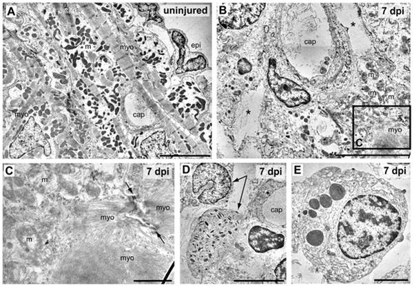
Fig. 3 Ultrastructural analysis of cryolesioned myocardium reveals cardiomyocyte cell death and an inflammatory response.
Electron micrographs of uninjured (A) and cryolesioned (7 dpi, B–E) zebrafish heart tissue. A normal organization of ventricular periphery with epicardal epithelium (epi) and subepicardial cardiomyocytes displaying well-organized striated myofilaments (myo) with Z lines and groups of electron dense mitochondria (m). A capillary (cap) is visible as well. (B) At 7 dpi, the lesioned area displays cellular debris and large tissue gaps (*) around a small capillary. In the lower right corner: cardiomyocyte with damaged mitochondria (m) and disorganised myofilaments (myo). (C) Magnification of the boxed area in B. A cryoinjured cardiomyocyte with disorganised mitochondria (m), myofilaments, and a loosened/ill defined intercalated disc (arrows) is shown. (D, E) Granulocytes in the wound area. (D) Heterophil granulocytes (arrows), the upper one with a peripheral, nonsegmented nucleus, the lower one with characteristic, cigar-shaped cytoplasmic granules. (E) Eosinophil granulocyte. Scale bars: 5 μm in A, B, and D, 1 μm in C, and 2 μm in E.