
Fig. s2
- ID
- ZDB-IMAGE-110517-6
- Publication
- Hicken et al., 2011 - Sublethal exposure to crude oil during embryonic development alters cardiac morphology and reduces aerobic capacity in adult fish
- All Figures
- Figures for Hicken et al., 2011
|
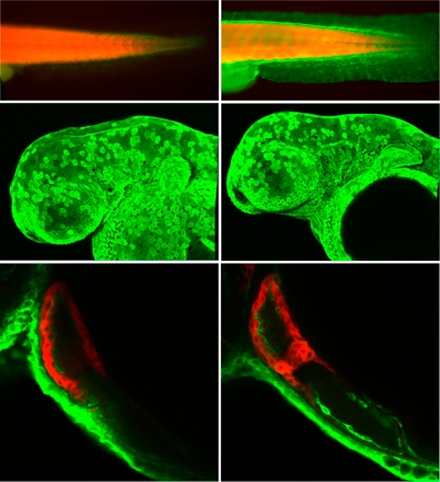
Fig. s2
CYP1A immunofluorescence in embryos exposed to gravel effluents. Embryos were exposed from 4 to 48 hpf in either clean (A) or oiled-gravel effluent (B–F), fixed and processed for whole-mount CYP1A (green) and myosin heavy chain (red) immunofluorescence as described under Materials and Methods. The myosin heavy chain antibody recognizes both skeletal and cardiac muscle and serves as an internal control for permeabilization. Images in A and B are epifluorescent, C and D are confocal stack projections, and E and F are confocal optical sections. Embryos from clutch 2 exposed to clean (A) or oiled (B) gravel effluent. (C and D) CYP1A immunofluorescence in the epidermal cells on the head of embryos from clutch 2 and clutch 3, respectively. Eye and pectoral fins (pf) are indicated. (E and F) CYP1A immunofluorescence in endocardial cells (arrowheads) of the cardiac atrium (a) and ventricle (v), and epithelium (arrows) over the pericardial region. (Scale bars: 100 μm.)