
|
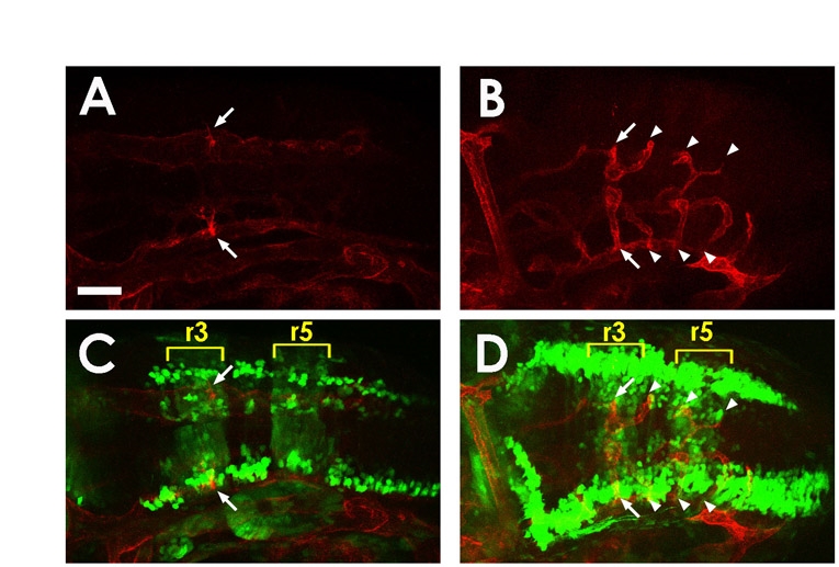
Fig. S2
Central arteries project through the center of the rhombomeres. (A-D) Confocal images of the hindbrain from a single Tg(kdrl:mCherry-CAAX)y171; Tg(pax2a:GFP)e1 double-transgenic embryo at 30 hpf (A,C) and 48 hpf (B,D), showing mCherry-positive blood vessels (A,B) or mCherry-positive blood vessels plus GFP-positive rhombomeres 3 and 5 (r3 and r5) and hindbrain neurons (C,D). The first pair of CtAs (white arrows) project dorsally through the middle of r3 (yellow brackets show r3 and r5), eventually linking directly with the BA. More posterior CtAs can be seen projecting through the center of r4, r5 and r6 (white arrowheads). Dorsal-lateral view, rostral to the left. Scale bar: 50 μm.