
Fig. 7
- ID
- ZDB-IMAGE-110429-17
- Publication
- Fujita et al., 2011 - Assembly and patterning of the vascular network of the vertebrate hindbrain
- All Figures
- Figures for Fujita et al., 2011
|
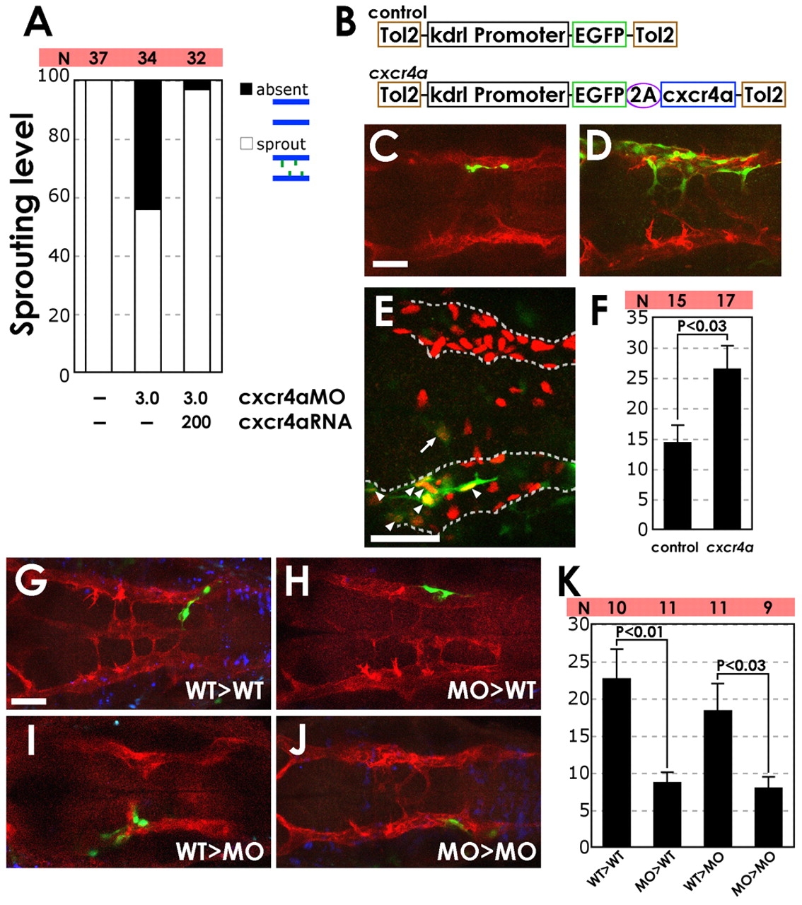
Fig. 7
Expression of cxcr4a promotes PHBC sprouting and medial migration in cxcr4a MO-injected animals. (A) Co-injection of 200 pg mRNA encoding wild-type cxcr4a alleviates the PHBC sprouting defect in zebrafish injected with 3 ng cxcr4a-MO2. The percentage of 30 hpf Tg(fli1a:EGFP)y1 animals showing endothelial cells migrating medially from the PHBC. The number of embryos analyzed for each injection is shown (N). (B) The Tol2 transposon cassettes of the control (kdrl:egfp) and cxcr4a (kdrl:egfp-2A-cxcr4a) expression vectors; the latter includes the 2A peptide sequence (see Materials and methods). (C,D) Confocal images of the hindbrain vasculature in 36 hpf cxcr4a-MO2-injected Tg(kdrl:mCherry-CAAX)y171 embryos co-injected with either control egfp transgene (C) or with a cxcr4a and egfp co-expression transgene (D). Dorsal view, rostral to the left. (E) Sample confocal image of the hindbrain vasculature in a 36 hpf Tg(kdrl:nls-mCherry)y173 embryo (with red fluorescent endothelial nuclei) injected with cxcr4a-MO2 and control kdrl:egfp transgene (green fluorescent endothelial cell bodies). In this animal, one EGFP-expressing endothelial cell (arrow) has migrated medially from the PHBC, whereas six EGFP-expressing endothelial cells (arrowheads) are present within the PHBC (outlined by a dashed line). Dorsal view, rostral to the left. (F) Quantification (percentage) of the medial migration tendency of transgene-expressing cells. The number of medially migrating EGFP-expressing endothelial cells (one in the example E) out of the total number of EGFP-expressing hindbrain endothelial cells (seven in the example E) was determined in 15 control (kdrl:egfp) and 17 cxcr4a (kdrl:egfp-2A-cxcr4a) animals. The s.e.m. is 2.9 for the control group and 3.9 for the cxcr4a group. The variability range of the two groups was comparable (P>0.1, F-test), and a t-test shows that the two groups are statistically different (P<0.03). (G-J) Confocal images of the hindbrain vasculature in Tg(kdrl:mCherry-CAAX)y171 transgenic host embryos from either wild type (WT) to WT (control MO-injected donor to control MO-injected host) (G), MO to WT (cxcr4a-MO2-injected donor to control MO-injected host) (H), WT to MO (I) or MO to MO (J) transplants. Dorsal view, rostral to the left. Transplanted cells were labeled with Dextran Blue. Transplanted vascular endothelial cells also express EGFP because Tg(fli1a:EGFP)y1 embryos were used as donors. (K) Quantification (percentage) of the medial migration tendency of transplanted cells. The number of medially migrating EGFP-expressing endothelial cells out of the total number of EGFP-expressing hindbrain endothelial cells was determined in ten (WT to WT), 11 (MO to WT), 11 (WT to MO) and nine (MO to MO) injected animals. Average percentages of medially migrated transplanted endothelial cells and s.e.m. were calculated for all groups. Scale bars: 50 μm.