
|
Fig. 1
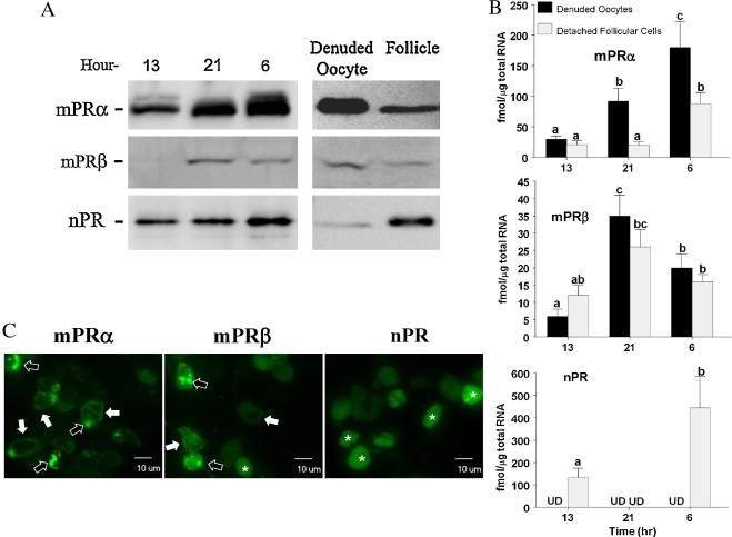
Expressions of membrane progestin receptor α (mPRα), mPRβ and nuclear progestin receptor (nPR or Pgr) in zebrafish oocytes and transfected human embryonic kidney 293 cell line (HEK293). (A) Representative Western analyses of mPRα, mPRβ and nPR proteins in follicle-enclosed stage IV oocytes (>650 μm) sampled at 13:00 (after spawning), 21:00 (evening), or 6:00 h (before spawning). The same analysis was also repeated in denuded oocytes and detached follicular cell layers collected at 6:00 (right panel in A). (B) Changes of mPRα, mPRβ, and nPR transcripts in detached follicular layers and denuded stage IV oocytes (n = 8) analyzed by real-time quantitative PCR (qRT-PCR). Different letters above the error bars indicate that those groups are significantly different from each other at P < 0.05. UD: under detection limit. (C) Localization of zebrafish mPRα-EGFP, mPRβ-EGFP, and nPR-EGFP proteins in transfected HEK293 cells. White arrows indicate plasma membrane localization; black arrows indicate intracellular compartment localization; and asterisks indicate cytoplasmic/nuclear localization. Each experiment was repeated at least three times.
Reprinted from Molecular and Cellular Endocrinology, 337(1-2), Hanna, R.N., and Zhu, Y., Controls of meiotic signaling by membrane or nuclear progestin receptor in zebrafish follicle-enclosed oocytes, 80-8, Copyright (2011) with permission from Elsevier. Full text @ Mol. Cell. Endocrinol.