
|
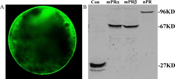
Fig. 3
(A) Expression of mPRα-GFP fusion proteins in a representative oocyte microinjected with the transgenic transcript. Follicular cells were removed for better imaging under a fluorescent microscope; (B) Western analyses of expressions of transgenic proteins for membrane progestin receptor α (mPRα), mPRβ, or nuclear progestin receptor (nPR) in protein extractions of purified plasma membrane (for mPRs) or cytosolic fraction (for nPR) from stage IV zebrafish oocytes. These oocytes were microinjected ex vivo with fused transcripts generated by fusing mPRα, mPRβ, or nPR in front of enhanced green fluorescent protein. Samples were collected following 5-h incubation in a culture medium. Representative results are shown as positive reactions (bands) to an anti-GFP antibody Con: control oocytes injected with a GFP carrier plasmid.
Reprinted from Molecular and Cellular Endocrinology, 337(1-2), Hanna, R.N., and Zhu, Y., Controls of meiotic signaling by membrane or nuclear progestin receptor in zebrafish follicle-enclosed oocytes, 80-8, Copyright (2011) with permission from Elsevier. Full text @ Mol. Cell. Endocrinol.