
Fig. S6
- ID
- ZDB-IMAGE-110321-25
- Genes
- Publication
- Wu et al., 2011 - SNW1 Is a Critical Regulator of Spatial BMP Activity, Neural Plate Border Formation, and Neural Crest Specification in Vertebrate Embryos
- All Figures
- Figures for Wu et al., 2011
|
Fig. S6
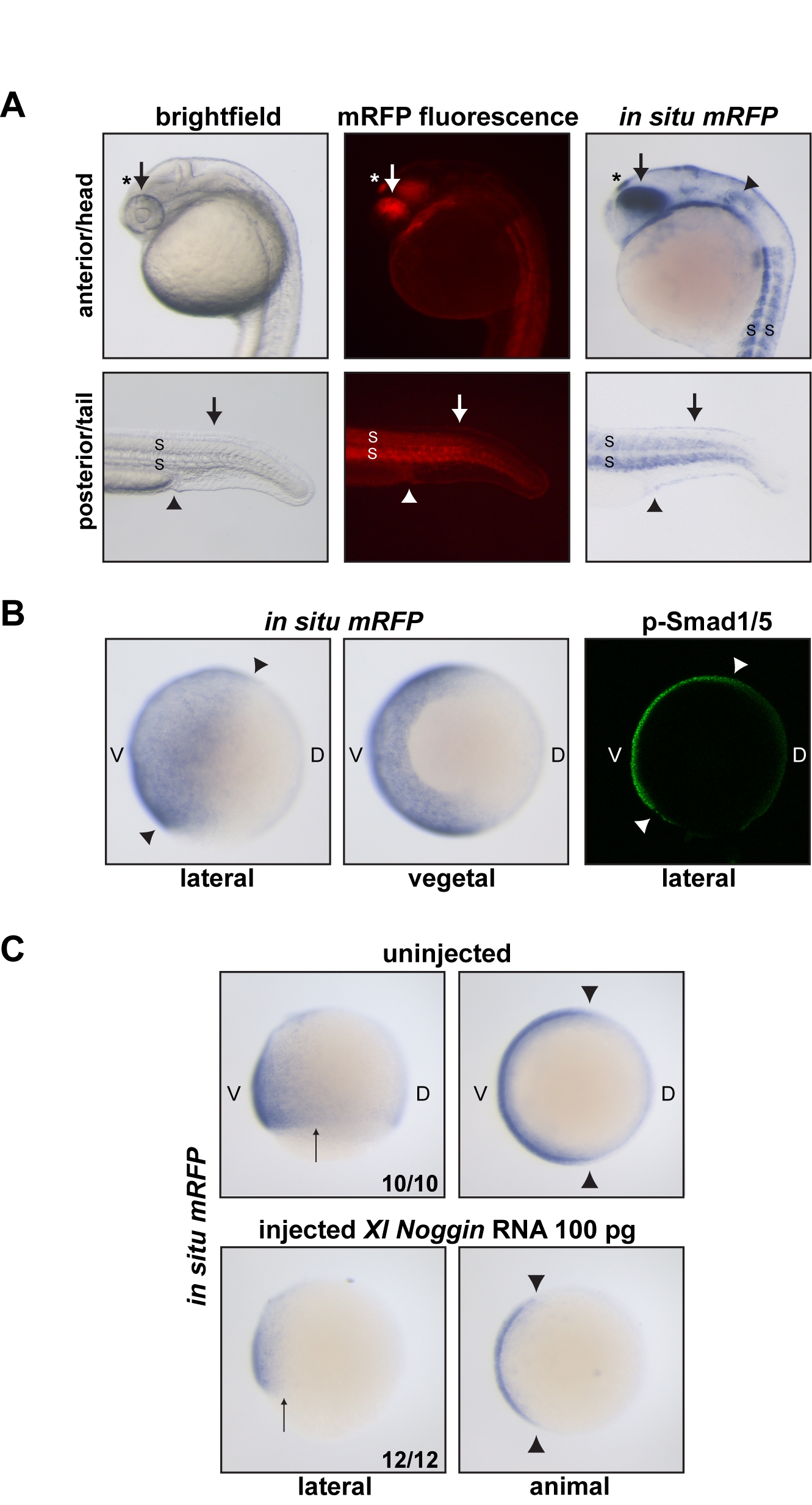
The BRE-mRFP transgenic zebrafish line is a reporter for BMP activity.
A) 24-h transgenic embryos imaged using bright field or fluorescence microscopy, or processed for in situ hybridization using a probe against mRFP. mRFP protein or mRNA is indicative of BMP activity. Indeed, in the anterior/head region, mRFP is detected in known sites of BMP expression and/or activity such as the dorsal retina and lens (arrow; [79]) and the otic placode (arrowhead; [80]). mRFP is also strongly detected in the epiphysis/pineal gland, consistent with data obtained in Xenopus (asterisk; [81]). In the posterior/tail region, mRFP as a readout of BMP signaling is present for instance in the dorsal ectoderm, where BMP ligands are expressed (arrow; [82]), the cloaca (arrowhead; [83]), and the somites (S; [82]). (B) mRFP in situ hybridization on transgenic embryos at 85% epiboly. mRFP transcripts are detected in the ventral ectoderm and the ventral lateral plate and intermediate mesoderm, where BMP signaling is known to be active [26]. The mRFP pattern is consistent with p-Smad1/5 staining at the same stage (arrowheads; see also [4]). (C) Noggin overexpression inhibits mRFP transcription downstream of the BRE promoter. As expected, injection of X. laevis Noggin mRNA results in reduced BMP signaling [26], which leads to lower amounts of mRFP transcripts in 70% epiboly embryos. Arrows and arrowhead indicate the extent of the mRFP expression domain for comparison between uninjected and injected embryos. In (B) and (C), V, ventral; D, dorsal. The number of embryos out of the total analyzed that showed the presented staining pattern is given.