Image
Figure Caption
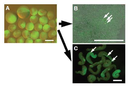
Fig. 1 Visualized green fluorescent protein (GFP)-positive cells obtained using the dissociated blastomere (DB) and cultured embryoid (CE) methods. (A) Isolation of blastodiscs at the late-blastula stage from embryos injected with GFP-nos1-3′UTR strand-capped mRNA. Large embryos were blastulae before operation, whereas small ones were isolated blastodiscs. (B) DBs cultured for 1 day in a dissociated condition in sodium citrate solution. (C) Embryoid from an isolated blastodisc cultured for 1 day in Ringer’s solution. Arrows indicate GFPpositive cells (primordial germ cells). Bars, 500 μm.
Acknowledgments
This image is the copyrighted work of the attributed author or publisher, and
ZFIN has permission only to display this image to its users.
Additional permissions should be obtained from the applicable author or publisher of the image.
Full text @ Int. J. Dev. Biol.

