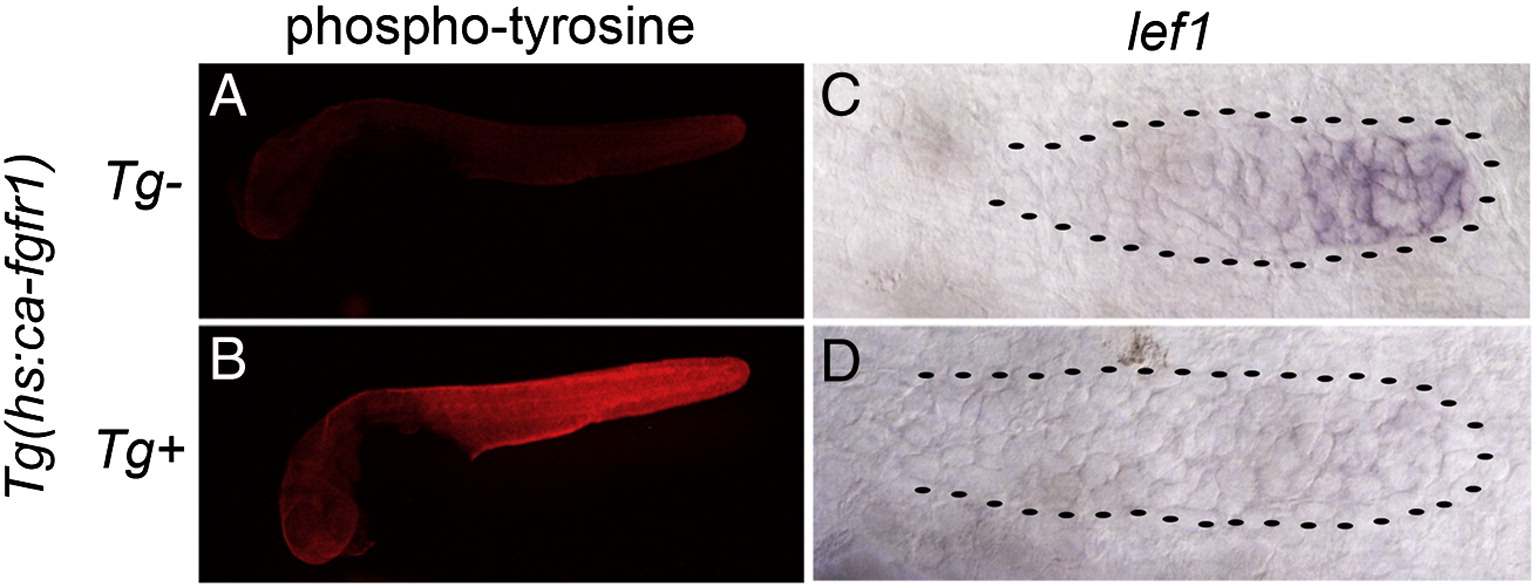
Fig. S3 Heatshock induction of ca-fgfr1 stimulates Fgf signaling in the primordium. (A,B) Whole mount phospho-tyrosine antibody staining at 32 hpf, six hours post heatshock. Embryos were mounted and photographed together to accurately display intensity difference. (A) Non-transgenic sibs express phospho-tyrosine broadly. (B) Embryos harboring the Tg(hs:ca-fgfr1) transgene show much more intense phospho-tyrosine staining demonstrating that the transgene activates Fgf signaling throughout the animal. (C,D) In situ hybridization with the Wnt/β-catenin pathway target lef1 in the primordium at 32 hpf six hours post heatshock. (C) In 32 hpf non-transgenic sibs lef1 is expressed in the leading zone of the primordium. (D) Induction of ca-fgfr1 leads to a loss of Wnt/β-catenin signaling in the primordium.
Reprinted from Developmental Biology, 349(2), Aman, A., Nguyen, M., and Piotrowski, T., Wnt/β-catenin dependent cell proliferation underlies segmented lateral line morphogenesis, 470-482, Copyright (2011) with permission from Elsevier. Full text @ Dev. Biol.

