
Fig. S3
- ID
- ZDB-IMAGE-110218-8
- Publication
- Barrios et al., 2003 - Eph/Ephrin signaling regulates the mesenchymal-to-epithelial transition of the paraxial mesoderm during somite morphogenesis
- All Figures
- Figures for Barrios et al., 2003
|
Fig. S3
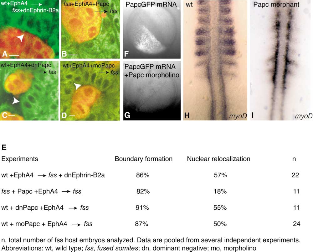
Epithelialization of Host Cells May Be Independent of Ephrin Reverse Signaling and Independent of Cell-Nonautonomous Activity of Papc
(A–D) Confocal images showing Bodipy 505-515-labeled somitic mesoderm of fss-/- host embryos (green) containing rhodamine dextranlabeled donor cells (red). Donor cells are wild-type cells expressing (A) EphA4, (B) fss-/- cells expressing EphA4 and Papc, (C) wild-type cells expressing EphA4 and dominant-negative Papc, or (D) wild-type cells expressing EphA4 and containing Papc morpholino. In (A), fss-/- host cells are expressing truncated, dominant-negative Ephrin-B2a-eGFP. The arrows point to basally localized nuclei of fss-/- host cells at boundaries created between donor and host cells. The scale bar represents 5 μm.
(E) Summary of experiments investigating the role of Ephrin-B2a and Papc upon epithelialization.
(F and G) Controls for the use of the papc morpholino (see the Supplemental Experimental Procedures).
(H and I) Dorsal views of somitic mesoderm of a wild-type embryo and an embryo injected with papc morpholino. Somite boundaries are not properly formed, and myoD expression is downregulated in the somites of Papc morphant embryos.