
|
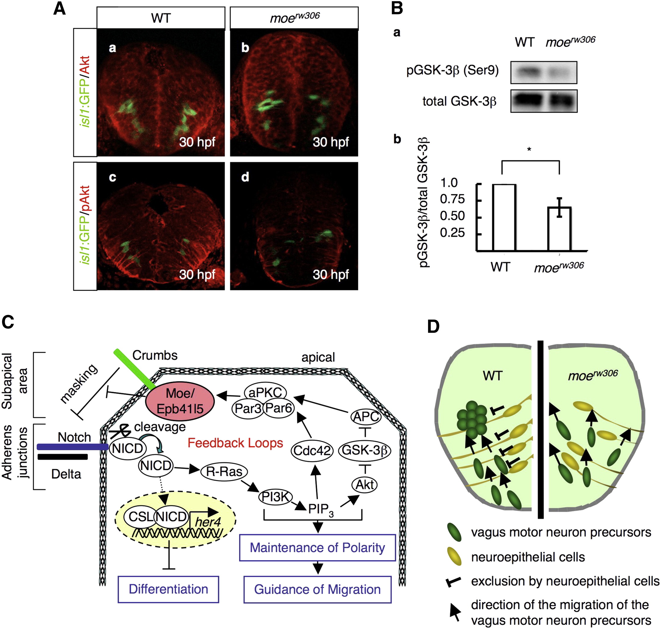
Fig. 8 Phosphorylation Levels of Akt and GSK-3β Are Reduced in the moerw306 Mutant
(A) Immunohistochemical detection of total Akt (a and b) and phospho-Akt (pAkt; c and d) in the WT (a and c) and moerw306 mutant (b and d) embryos at 30 hpf. The vagus motor neurons are also shown (green). Cross-sectional views, dorsal to the top.
(B) (a) Western blotting with anti-total GSK-3β and anti-phospho-GSK-3β (pGSK-3β) antibodies of the WT and moerw306 mutant lysates from 30–34-hpf embryos. (b) The relative intensities of the pGSK-3β bands were quantified with the ImageJ software and normalized to those of the total GSK-3β bands. Data shown are mean ± SEM; *p < 0.05.
(C) Schematic of the hypothetical feedback loop, which may function in the maintenance of neuroepithelial polarity.
(D) A model for neuroepithelial cell guidance of the migration of the vagus motor neuron precursors. Cross-sectional view, dorsal to the top.