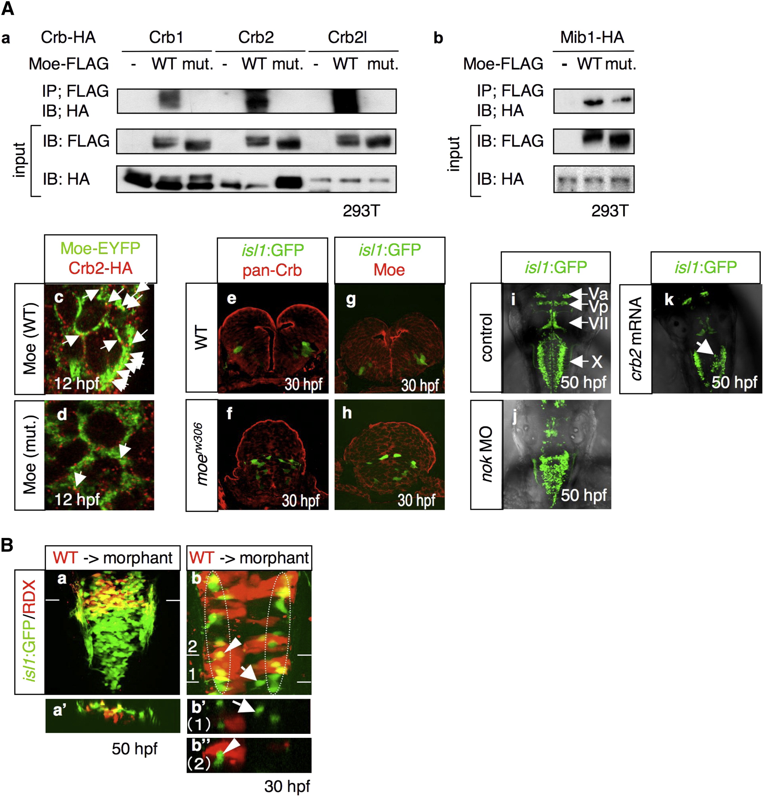
Fig. 2 Moe and the Crb Family Proteins Forms a Complex that Is Localized to the Apical Surface of Neuroepithelial Cells
(A) (a) Coimmunoprecipitation of Moe-FLAG with Crb1-HA, Crb2-HA, and Crb2l-HA. 293T cells were transfected with a plasmid that encodes HA-tagged Crb1, Crb2, or Crb2l, together with the mock plasmid (-), or the plasmids that encode FLAG-tagged WT Moe (WT) and L221R-type Moe (mut.). (b) Coimmunoprecipitation of Moe-FLAG and Mib-HA. The following abbreviations are used: IP, immunoprecipitation; IB, immunoblot. (c and d) Localization of Crb2-HA (c and d; red), WT Moe-EYFP (c; WT; green), and L221R-type Moe-EYFP (d; mut.; green) in WT neuroectodermal cells at 12 hpf. Arrows indicate the colocalization of Crb2-HA and Moe-EYFP. The mRNAs for these proteins were injected at 60 μg/ml. (e–h) Localization of the Crb family proteins (e and f; red) and Moe (g and h; red) in the WT (e and g) and moerw306 mutant (f and h) embryos at 30 hpf. The vagus motor neurons appear in green (cross-sectional view, dorsal to the top). (i–k) Dorsal view of the embryos injected with the 5-mis-pair control MO (i; 2.5 mg/ml), nok MO (j; 2.5 mg/ml), and crb2 mRNA (k; 500 μg/ml) at 50 hpf. The arrow in (k) indicates the aberrant positioning of the vagus motor neurons.
(B) (a and a′) The WT vagus motor neuron progenitors (yellow) were transplanted into the moe morphant hindbrain. The images taken at 50 hpf show the dorsal view (a, rostral to the top) and cross-sectional view (a′; dorsal to the top) at the level of the line shown in (a). (b, b′, and b″) WT cells (red) were transplanted into the dorsomedial region of the moe morphant hindbrain. In this case, at 30 hpf, the morphant vagus motor neuron progenitors have migrated to the correct locations (enclosed by dotted-line), while those neurons that are not surrounded by the WT cells are positioned medially (arrows in b and b′). The images in b′ and b″ are the cross-sectional views (dorsal to the top) of the dorsal views of b (rostral to the top) at the level of line 1 and 2, respectively. Note that some of the morphant-derived vagus motor neurons appear in yellow owing to the fact that (b) shows stacked images. However, they actually appear in green in the cross-section (arrowheads in b and b″). RDX is an abbreviation of rhodamine-dextran.

