
Fig. 6
- ID
- ZDB-IMAGE-110214-39
- Antibodies
- Publication
- Ravanelli et al., 2011 - The Actin Nucleator Cordon-bleu is Required for Development of Motile Cilia in Zebrafish
- All Figures
- Figures for Ravanelli et al., 2011
|
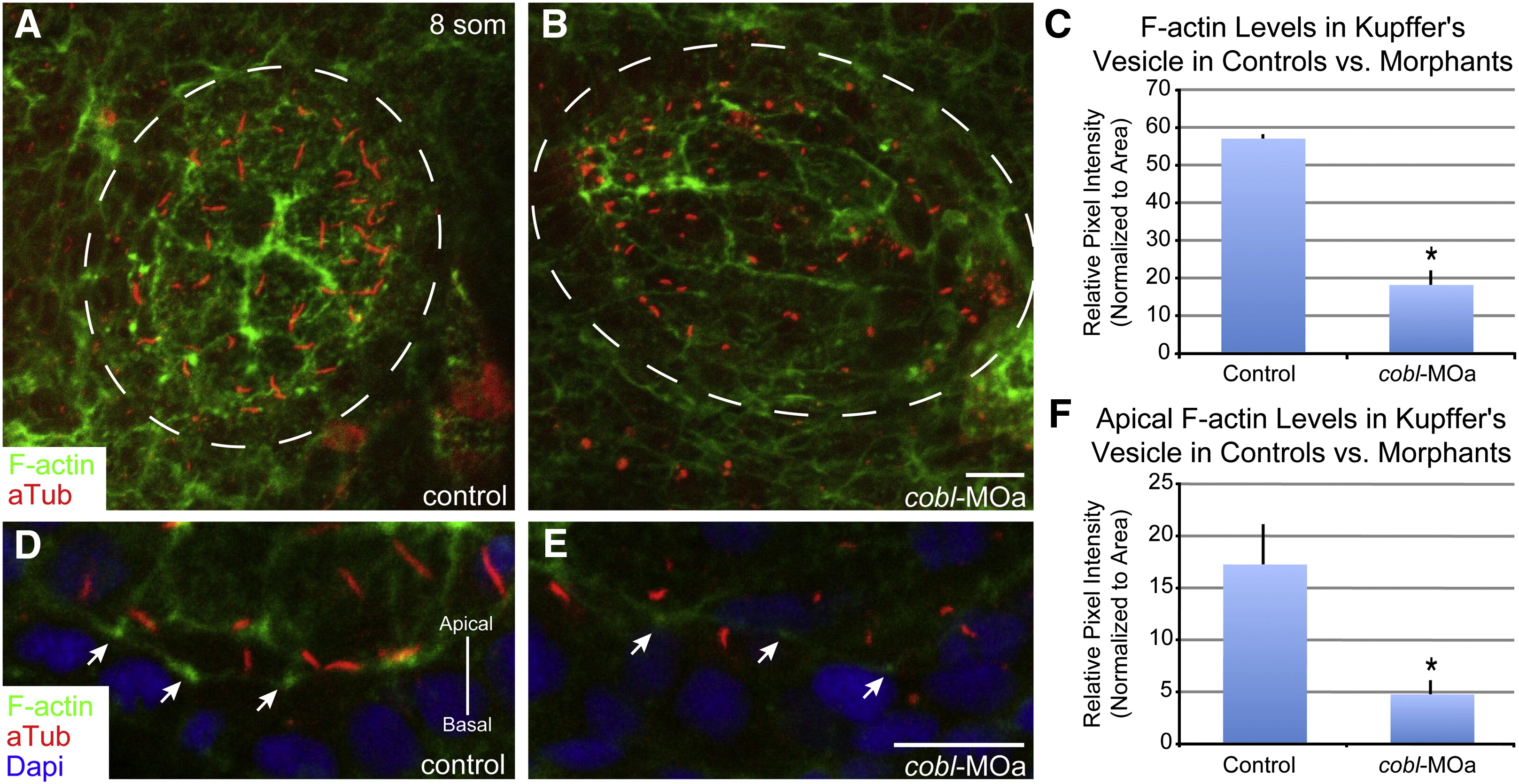
Fig. 6 Reduction of Cobl causes a reduction of apical F-actin in Kupffer′s vesicle.
(A–B) Maximum intensity projection of 0.8 μm confocal z-stacks of Kupffer′s vesicle, stained for cilia and actin. cobl morphant embryos with short cilia show deficits in the amount of F-actin staining by phalloidin. (C) Bar graph of pixel intensity of F-actin in Kupffer′s vesicle in controls vs. cobl morphants. Morphant embryos exhibit a significant decrease in the amount of F-actin. (D–E) Confocal images of cells in Kupffer′s vesicle showing cilia and F-actin. (F) Bar graph of pixel intensity of apical F-actin in Kupffer′s vesicle cells in controls vs. cobl morphants. Morphant embryos exhibit a significant decrease in the amount of F-actin present at the apical surface of cells. Dashed lines outline Kupffer′s vesicle. Arrows demark cortical F-actin. F-actin (phalloidin), cilia (anti-acetylated tubulin), DNA (DAPI). n values: control = 12, MOa = 7. p values: C = 0.0012*, F = 0.0283*. Error bars represent one standard error from the mean. Scale bar represents 10 μm.
Reprinted from Developmental Biology, 350(1), Ravanelli, A.M., and Klingensmith, J., The Actin Nucleator Cordon-bleu is Required for Development of Motile Cilia in Zebrafish, 101-111, Copyright (2011) with permission from Elsevier. Full text @ Dev. Biol.